Using AI for Atypical Rashes: How Rash Detector Helps Identify Unusual Skin Conditions
Using AI for atypical rashes can speed diagnosis and guide care. Learn how Rash Detector analyzes photos to spot unusual skin issues accurately.
Estimated reading time: 18 min
Key Takeaways
- AI tools analyze color, shape, texture, and pattern to help identify atypical rashes from photos.
- Rash Detector uses advanced AI to assist in recognizing eczema, psoriasis, fungal infections, viral rashes, Lyme disease, and more.
- Photo quality—lighting, focus, and angles—greatly impacts AI accuracy for rash detection.
- AI can aid triage and early assessment but cannot replace dermatologist consultations for complex or severe cases.
- Safety guidelines and privacy protections are important when uploading rash images to AI apps.
Table of Contents
- Section 1: Understanding AI in Atypical Rash Detection
- Section 2: Visual Features AI Uses to Classify Rashes
- Section 3: Common Rash Categories AI Can Recognize
- Section 4: Accuracy, Limitations, and When to See a Doctor
- Section 5: Tips for Taking Photos That Improve AI Diagnosis
- Section 6: Privacy, Safety, and Medical Disclaimer for AI Rash Apps
Section 1: Understanding AI in Atypical Rash Detection
What Is AI-Based Rash Detection?
Artificial intelligence (AI) for atypical rashes involves using advanced machine learning (ML) models trained on extensive datasets comprising thousands of labeled skin images. These images cover a wide spectrum of dermatologic conditions, including some rare and unusual rashes that may be challenging to diagnose clinically. By analyzing photographs uploaded by users, AI systems assess visual cues such as color, shape, texture, and distribution patterns to generate a list of possible diagnoses and recommend appropriate next steps.
Apps like Rash Detector provide user-friendly interfaces enabling individuals to upload multiple high-resolution images of their rash. The AI then processes these images quickly, offering real-time feedback that can help users better understand the nature of their skin condition. This technology may be particularly helpful for those without immediate access to dermatologists or in regions where specialist care is limited.
How AI Models Learn to Identify Rashes
AI models, particularly convolutional neural networks (CNNs), learn to identify rashes through a training process using labeled datasets containing thousands to hundreds of thousands of images. These datasets include images of common and atypical rashes across diverse skin tones, ages, and body locations to improve model generalizability. Training involves iterative adjustments of the model’s parameters to minimize error in classification tasks.
The AI learns to detect subtle differences in features that may be difficult to distinguish for the untrained eye. For example, it can discern differences between the fine scaling of psoriasis plaques and the coarser, dry patches of eczema, or detect the central clearing characteristic of fungal infections like ringworm. This pattern recognition extends beyond mere color or shape to complex interplays of visual information, including micro-texture and lesion distribution.
Continual model refinement occurs as new images and clinical feedback are incorporated, improving accuracy over time. However, the AI’s performance still depends heavily on the diversity and quality of training data, which is why rare or novel presentations can pose challenges.
Benefits of AI for Atypical Rashes
- Speed: AI provides immediate preliminary assessments, which can be useful for patients needing guidance on whether to seek emergency care or routine dermatologic evaluation.
- Accessibility: AI-powered rash detection can help increase access to dermatology expertise, especially in underserved areas or for patients with mobility constraints.
- Educational: The app can offer explanations about possible diagnoses and general skin health tips, empowering users to better understand their condition.
- Support for Clinicians: In clinical settings, AI tools may assist dermatologists by highlighting suspicious features or suggesting differential diagnoses, potentially improving diagnostic workflow.
- Data Collection: Aggregated anonymized data can support epidemiological research on rash prevalence and presentation patterns.
Section 2: Visual Features AI Uses to Classify Rashes
Color Patterns
Color analysis is a foundational aspect of AI rash classification. The AI examines not only the dominant color but also subtle variations and gradients within lesions. For instance:
- Redness (Erythema): Common in inflammatory rashes like eczema or contact dermatitis; intensity may correlate with severity.
- Purpura or Purple: May indicate vascular inflammation or bleeding under the skin, seen in vasculitis or certain drug reactions.
- Brown or Hyperpigmentation: Often appears in chronic rashes or post-inflammatory changes.
- Blanching: AI may infer blanching behavior (whether redness fades when pressed) indirectly by color distribution and saturation patterns.
Precise color calibration is important, which is why consistent lighting and minimal image editing improve AI performance.
Shape and Borders
The morphology of individual lesions and their borders provide critical diagnostic clues. The AI categorizes shapes into:
- Round or Oval Lesions: Seen in fungal infections like ringworm or in fixed drug eruptions.
- Linear Lesions: May suggest dermatitis along scratch lines or herpes zoster following nerve dermatomes.
- Irregular Shapes: Typical of eczema or some viral exanthems.
Border sharpness is also analyzed:
- Well-demarcated borders: Common in psoriasis plaques or fungal infections.
- Ill-defined borders: Eczema and some allergic reactions tend to have diffuse edges.
Additionally, the AI evaluates lesion clustering and spacing patterns, which can help differentiate between grouped vesicles in herpes and scattered papules in viral exanthems.
Texture and Surface Features
Texture assessment involves detecting surface changes such as scaling, crusting, vesicles, or pustules. The AI compares pixel intensity variations and reflective properties:
- Scaling: Fine white or silver flakes, typical of psoriasis, contrast with the dry, flaky skin of eczema.
- Blistering: Presence of vesicles or bullae can indicate herpes infections, autoimmune blistering diseases, or contact dermatitis.
- Crusting and Oozing: Suggest secondary infection or exudative phases of dermatitis.
Advanced AI algorithms also analyze three-dimensional cues from shadows and highlights to better infer texture depth.
Distribution and Pattern
AI examines spatial arrangement across the body or photographed area, which is important for diagnosis:
- Localized vs. Generalized: Localized rashes might indicate contact dermatitis or insect bites, while generalized rashes may suggest viral exanthems or systemic conditions.
- Symmetry: Symmetrical distribution is common in eczema and psoriasis, whereas asymmetry may indicate infections or allergic reactions.
- Following Dermatomes or Nerve Paths: Characteristic of herpes zoster.
- Annular or Target Patterns: Seen in Lyme disease’s erythema migrans or erythema multiforme.
By integrating these spatial data with lesion characteristics, AI aims to improve diagnostic precision.
Section 3: Common Rash Categories AI Can Recognize
Eczema and Psoriasis
Both eczema (atopic dermatitis) and psoriasis are chronic inflammatory skin diseases with distinct features:
- Eczema: AI identifies patchy erythema with fine scaling, dryness, and often excoriations due to itching. Lesions are commonly found in flexural areas (behind knees, inside elbows) and may feature oozing when acute.
- Psoriasis: Characterized by sharply demarcated plaques with thick, silvery scales predominantly on extensor surfaces like elbows and knees. AI detects the unique scale texture and border definition, differentiating it from eczema.
AI may sometimes estimate disease severity by analyzing lesion size and scaling intensity, which could aid in monitoring treatment response.
Fungal and Bacterial Infections
Fungal infections such as dermatophytosis (ringworm) present with circular or oval lesions having central clearing and raised, scaly borders. Candida infections often appear as moist, erythematous patches in intertriginous areas. AI recognizes these patterns through shape and texture cues.
Bacterial infections may manifest as impetigo with honey-colored crusts or folliculitis presenting with pustules. AI identification relies on recognizing pustular lesions and crusting patterns that help distinguish bacterial from fungal or inflammatory rashes.
Additionally, AI can flag signs of secondary infection complicating chronic rashes, assisting users to seek timely medical care.
Viral Rashes
Viral exanthems exhibit characteristic features and progression:
- Measles: Maculopapular rash starting on the face and spreading downward, often with Koplik spots inside the mouth.
- Chickenpox: Vesicular rash at different stages—papules, vesicles, crusts—distributed on the trunk and face.
- Herpes Simplex Virus: Clustered vesicles on erythematous base, often localized around the mouth or genitals.
AI models trained on temporal progression and lesion morphology may suggest viral etiologies by analyzing multiple images over time or identifying lesion types and distribution.
Lyme Disease Rash (Erythema Migrans)
The hallmark bull’s-eye rash of early Lyme disease consists of an expanding red patch with central clearing. AI trained on documented cases can recognize this pattern despite its variability in size and color intensity. Early detection is important as Lyme disease requires prompt antibiotic treatment to prevent systemic complications.
AI can also assist in differentiating erythema migrans from similar-appearing rashes such as cellulitis or ringworm, helping users prioritize medical evaluation.
Section 4: Accuracy, Limitations, and When to See a Doctor
How Accurate Are AI Rash Detectors?
Published data on AI dermatology tools show promising results, with some AI systems achieving diagnostic accuracy rates around 80-90% for common rashes when compared with board-certified dermatologists. However, these results depend heavily on factors such as image quality, the diversity of training data, and the specific skin condition.
For atypical or rare presentations, AI confidence scores tend to be lower, reflecting uncertainty. Rash Detector displays these confidence metrics alongside suggested diagnoses to help users interpret results responsibly. Moreover, AI systems are continuously updated with new data to refine performance.
It is important to recognize that AI accuracy is complementary rather than definitive, serving primarily as a decision support tool.
Limitations of AI for Atypical Rashes
- Mixed or Overlapping Conditions: Patients with multiple concurrent skin diseases can present complex images that challenge AI classification.
- Underlying Systemic Disease: Some rashes are manifestations of internal conditions (e.g., lupus, vasculitis), requiring clinical and laboratory evaluations beyond visual assessment.
- Physical Examination: AI cannot palpate lesions to assess texture, tenderness, or induration, nor can it evaluate associated lymphadenopathy or systemic signs.
- Patient History and Symptoms: Clinical context is essential for diagnosis; AI lacks access to patient history, symptom chronology, and response to treatments.
- Varied Skin Tones: Despite efforts to include diverse data, AI may perform less accurately on underrepresented skin types due to color and texture differences.
When to Consult a Dermatologist
While AI can guide early triage, professional evaluation is essential in the following circumstances:
- Rapidly Spreading or Worsening Rash: May indicate infection or severe allergic reaction requiring urgent care.
- Painful, Blistering, or Bleeding Lesions: Could signify serious conditions like bullous diseases or necrotizing infections.
- Systemic Symptoms: Presence of fever, malaise, joint pain, or difficulty breathing alongside rash necessitates prompt medical attention.
- Non-Improvement After Initial Treatment: If rash persists or worsens despite following AI app recommendations or over-the-counter remedies.
- New or Unexplained Rashes in Children or Elderly: Vulnerable populations require careful assessment for infections or immune disorders.
In these situations, users should seek timely consultation with a board-certified dermatologist or healthcare provider for comprehensive diagnosis and management.
Remember, Rash Detector is intended as an adjunctive tool and not a substitute for professional medical advice.
Section 5: Tips for Taking Photos That Improve AI Diagnosis
Lighting and Focus
Optimal lighting is crucial to capture accurate color and texture details. Use natural daylight or well-diffused artificial lighting to avoid harsh shadows, glare, or color distortion. Avoid direct flash, which can cause reflections or wash out colors.
Maintain steady hands or use a tripod/stand to reduce blurring. Use your camera’s autofocus function by tapping the rash area on the screen to ensure sharp focus. Multiple photos may be necessary to capture clear images, especially in low-light conditions.
Multiple Angles and Surrounding Skin
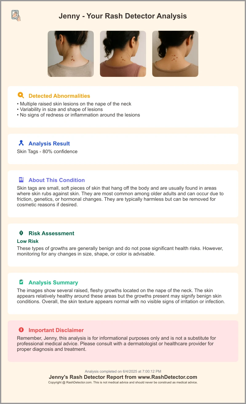
Upload at least three images to provide comprehensive information:
- Close-up: Shows detailed lesion morphology, including surface features and borders.
- Medium Distance: Captures the lesion in context with surrounding skin, revealing color contrast and scale.
- Wider View: Shows distribution pattern and relation to body landmarks.
Photographing surrounding unaffected skin aids AI in comparing lesion characteristics and enhances classification accuracy.
Consistent Background and Scale
Use a plain, neutral background such as a white or light-colored sheet to avoid distractions. Include an object of known size—a ruler, coin, or credit card—next to the rash to provide scale. This helps the AI estimate lesion dimensions, which can be diagnostically relevant.
Avoid applying photo filters, color adjustments, or cropping that may alter the true appearance of the rash. Authentic images yield more reliable AI assessments.
Section 6: Privacy, Safety, and Medical Disclaimer for AI Rash Apps
Data Privacy and Security
Rash Detector prioritizes user privacy by employing end-to-end encryption during image upload and data transmission. All images and associated metadata are stored securely on servers compliant with healthcare data protection standards such as HIPAA (in applicable regions) and GDPR.
User data is anonymized and utilized only for AI model improvement and research with explicit consent. The platform does not share personal health information with third parties without permission, maintaining strict confidentiality.
Medical Disclaimer and Safe Use
AI rash detection tools are designed to provide educational information and preliminary assessments, not definitive diagnoses. Users must understand that these apps are adjuncts to, not replacements for, professional healthcare evaluation.
Always consult a licensed healthcare provider or dermatologist for diagnosis, treatment planning, and follow-up care, especially if your rash is severe, persistent, or worsening.
Recognizing Red-Flag Symptoms
If your rash is accompanied by any of the following, seek emergency medical attention immediately instead of relying solely on AI analysis:
- Difficulty breathing or swallowing
- Swelling of the face, lips, or tongue
- High fever or chills
- Rapidly spreading skin redness or blistering
- Severe pain or signs of systemic infection
These symptoms may indicate life-threatening allergic reactions, infections, or systemic illnesses requiring urgent intervention.
Conclusion
AI-based analysis for atypical rashes represents a promising advancement in dermatologic care accessibility and early detection. By evaluating key visual features—color nuances, lesion shape and borders, texture variations, and distribution patterns—AI tools like Rash Detector provide users with valuable insights into potential skin conditions. This technology can empower individuals to make informed decisions about seeking medical care and support clinicians with diagnostic suggestions.
However, the effectiveness of AI depends significantly on high-quality images and user understanding of its limitations. It cannot replace comprehensive clinical evaluation, particularly for complex, severe, or systemic skin diseases. Following best practices for photographing rashes and adhering to privacy and safety guidelines helps ensure optimal AI performance and protects user data.
For those exploring AI-assisted dermatology, Rash Detector offers a secure, user-friendly platform to upload multiple photos and receive instant, AI-powered rash evaluations—an excellent first step toward managing atypical skin conditions responsibly and proactively.
FAQ
Q: Can AI identify an atypical rash from a photo?
A: AI trained on large, diverse datasets can assist in recognizing many atypical rashes by analyzing visual features such as color, shape, texture, and distribution. However, accuracy varies with how unusual or complex the rash is, and AI results should be interpreted as preliminary guidance.
Q: How accurate are AI rash detectors compared with dermatologists?
A: AI systems can approach dermatologist-level accuracy—often around 80-90% for common rashes—when provided with high-quality images. For rare, mixed, or atypical conditions, AI accuracy may be lower. Confidence scores provided with results help users understand diagnostic certainty.
Q: What kinds of rashes can AI recognize?
A: AI can assist in identifying a range of conditions including eczema, psoriasis, fungal and bacterial infections, viral rashes, and Lyme disease’s erythema migrans. Continuous model updates aim to expand this spectrum.
Q: When should I see a doctor instead of relying on an AI rash checker?
A: Consult a healthcare professional if your rash is spreading rapidly, painful, blistering, accompanied by systemic symptoms like fever, or does not improve after following initial care. Also, seek immediate emergency care for symptoms such as difficulty breathing or severe swelling.
Q: How do I take a good photo for AI rash analysis?
A: Use natural, diffuse lighting to avoid shadows; keep the rash area in sharp focus; take multiple photos from different angles and distances including surrounding skin; use a plain background and include a size reference like a ruler or coin; avoid filters or editing.
Q: Can AI tell the difference between eczema, psoriasis, and fungal rashes?
A: AI evaluates distinct visual features like scaling type, border definition, lesion shape, and distribution patterns to help differentiate these conditions with reasonable accuracy, supporting further medical evaluation.