Skin Checks by RNs: Essential Guide for Comprehensive Assessments
Learn how skin checks by RNs help detect rashes, pressure injuries, and suspicious lesions early for better patient care and safety.

Estimated reading time: 18 min
Key Takeaways
- Skin checks by RNs involve thorough inspection and palpation to identify rashes, lesions, and pressure injuries, utilizing systematic head-to-toe approaches.
- The ABCDE criteria provide guidance for evaluating suspicious moles to help identify lesions that may require further medical evaluation.
- High-risk patients may require more frequent skin assessments, often daily or per shift, with detailed documentation to monitor progression and aid interdisciplinary communication.
- Assessment includes evaluating color, temperature, moisture, turgor, edema, and skin integrity, with attention to subtle changes that may indicate systemic or localized issues.
- Early escalation to providers or dermatologists is important when abnormalities are found, and adjunctive tools like AI-driven apps can support clinical decision-making but do not replace professional evaluation.
Table of Contents
- Section 1: Fundamentals of Skin Checks by RNs
- Section 2: Full-Body Skin Assessment Components
- Section 3: Using ABCDE Criteria to Identify Suspicious Lesions
- Section 4: Differentiating Skin Cancer Screening and Routine Nursing Skin Checks
- Section 5: Recognizing Risk Factors for Frequent Skin Assessments
- Section 6: Pressure Injury Prevention and High-Risk Body Sites
- Section 7: Documentation and When to Escalate Findings
Conclusion
FAQ
Section 1: Fundamentals of Skin Checks by RNs
What Are Skin Checks by Registered Nurses?
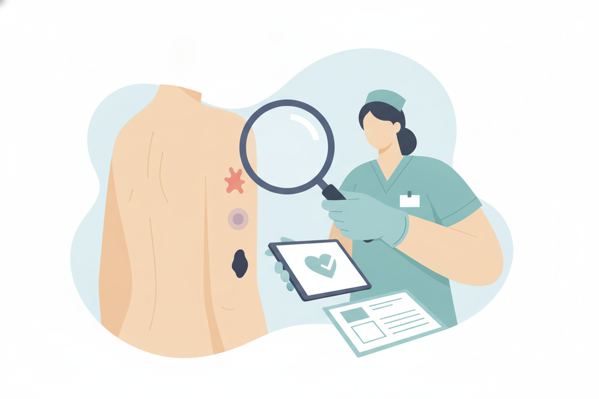
Skin checks by registered nurses (RNs) are systematic, methodical evaluations of a patient’s skin conducted as an integral part of nursing assessments in various care settings, including hospitals, long-term care facilities, outpatient clinics, and home care. These checks are important for identifying early signs of skin abnormalities such as rashes, pressure injuries (also known as pressure ulcers or bedsores), suspicious lesions, infections, and other dermatological conditions. The goal is to help ensure skin integrity, recognize potential complications early, and initiate timely interventions.
RNs employ a combination of inspection and palpation techniques to assess skin health. Inspection allows for visual identification of changes in skin appearance, while palpation provides tactile information about texture, temperature, moisture, and other physical characteristics that may not be evident visually. Together, these techniques enable nurses to detect subtle abnormalities that can be easily missed without careful examination.
Inspection and Palpation Techniques
Effective skin checks require a structured approach to inspection and palpation, ensuring all relevant parameters are evaluated systematically. Below is an explanation of key elements assessed during these techniques:
- Color: Nurses observe for variations from the patient’s baseline skin tone. Redness (erythema) may indicate inflammation or infection; pallor suggests anemia or poor perfusion; cyanosis (bluish discoloration) may indicate hypoxia; and jaundice (yellowing) may reflect liver dysfunction. For example, localized redness over a bony prominence may be an early sign of pressure injury.
- Temperature: Using the dorsum of the hand, RNs assess skin temperature bilaterally. A warm or hot area can signify infection, inflammation, or vascular congestion. Conversely, cool skin may suggest poor arterial blood flow or shock. Comparing symmetrical body parts enhances detection of abnormalities.
- Moisture: Skin moisture is evaluated by palpation and visual inspection. Excessive dryness can cause fissures or cracks, increasing infection risk, while excessive sweating (diaphoresis) might be related to systemic illness or medication effects. For instance, a dry scaly rash could indicate eczema or xerosis.
- Turgor: Skin turgor reflects hydration status and elasticity. Pinching the skin gently over a bony area (such as the forearm or clavicle) and observing how quickly it returns to normal provides insight into fluid balance. Poor turgor (slow return) can indicate dehydration, which is more common in elderly patients.
- Edema: Palpation helps identify the presence and extent of fluid accumulation in tissues. Pitting edema (indentation remaining after pressure) can suggest heart failure, renal insufficiency, or localized venous obstruction. Documenting the grade of edema (1+ to 4+) aids in tracking progression.
Importance of Early Detection
Early detection of skin abnormalities during nursing assessments is important to help prevent complications and optimize patient outcomes. For example, recognizing the onset of a drug-induced rash early allows for prompt discontinuation of the offending agent and initiation of appropriate treatment, potentially preventing progression to severe reactions.
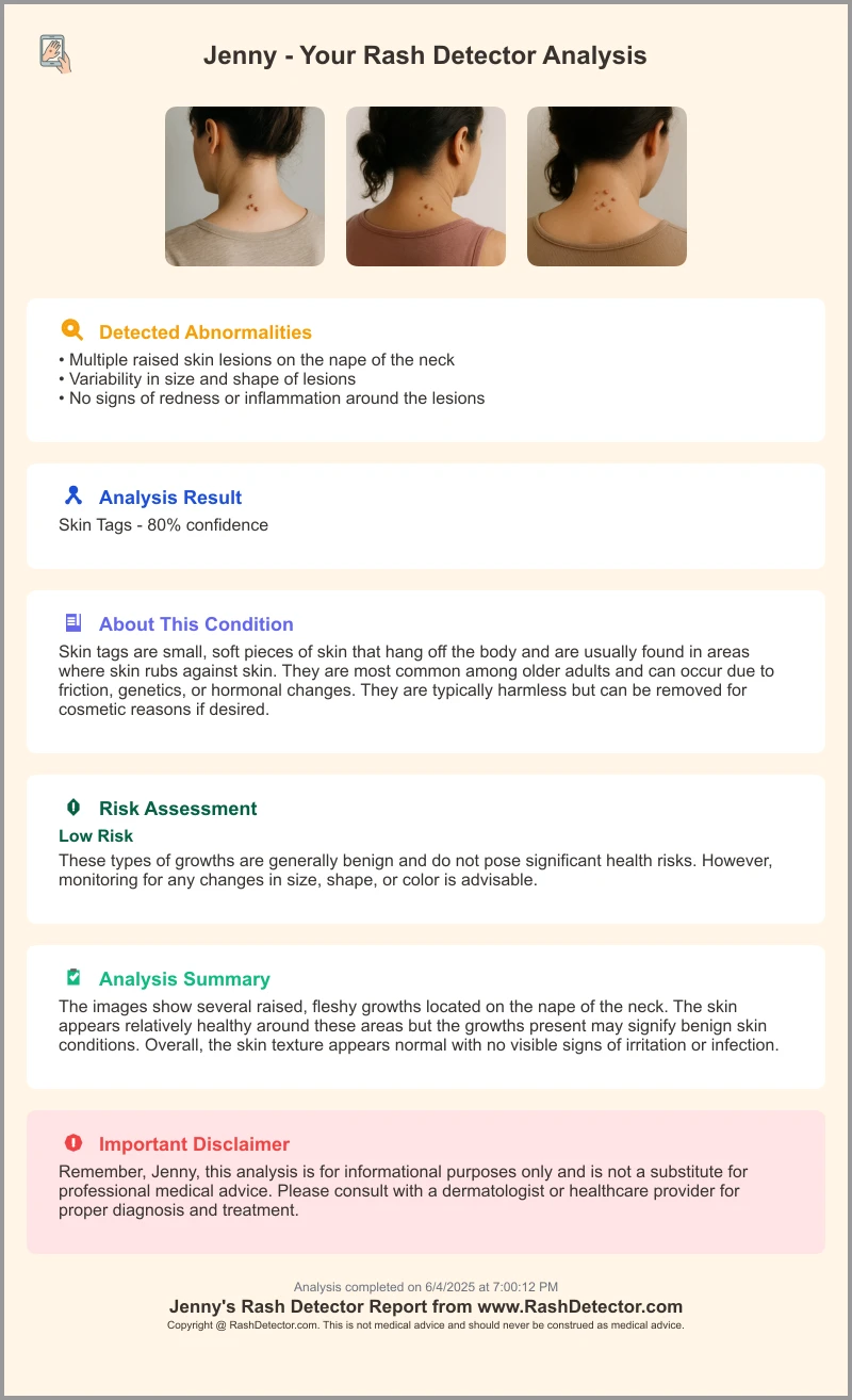
The Rash Detector app can be a useful tool in this regard, offering AI-driven analysis of rash images uploaded by clinicians. By providing diagnostic suggestions and severity assessments, the app may aid nurses and providers in making informed decisions, enhancing patient safety and care quality.
Moreover, early identification of suspicious lesions or pressure injuries enables timely intervention, which may reduce morbidity, hospital stays, and healthcare costs. RNs play a pivotal role in initiating this critical first step in skin health management.
Section 2: Full-Body Skin Assessment Components
Scope of a Comprehensive Skin Check
A comprehensive full-body skin check by an RN is a deliberate, head-to-toe examination that covers all skin surfaces, including often overlooked areas. This thorough approach helps ensure no abnormalities are missed, especially in patients with risk factors such as immobility, impaired sensation, or chronic illness.
The examination typically includes:
- Head and face, including the scalp and hairline
- Neck and torso, both anterior and posterior surfaces
- Upper and lower extremities, including palms, soles, and interdigital spaces
- Nails and cuticles
- Mucous membranes, including oral and genital areas
- All bony prominences and pressure points prone to skin breakdown
Key Areas to Examine
- Scalp: Inspection involves parting the hair to assess the scalp skin for lesions such as psoriasis plaques, seborrheic dermatitis, or signs of infection like folliculitis. Palpation can detect tenderness or raised nodules.
- Nails: Evaluate nail color, thickness, shape, and integrity. Changes like clubbing, brittle nails, onychomycosis (fungal infection), or splinter hemorrhages may indicate systemic conditions.
- Pressure Points: These include heels, sacrum, elbows, hips, occiput (back of the head), and malleoli. These sites are prone to pressure injuries due to prolonged pressure, especially in immobile patients. Look for redness, blanching, skin breakdown, or ulceration.
- Mucous Membranes: Oral mucosa may reveal dehydration (dryness), candidiasis (white patches), or petechiae (small hemorrhages). Genital mucosa assessment can detect infections or dermatological conditions such as lichen sclerosis.
Techniques for Effective Skin Checks
To maximize accuracy and patient comfort during skin assessments, RNs should:
- Use adequate lighting, preferably natural or bright artificial light, to enhance visualization of subtle color changes.
- Wear gloves to maintain hygiene and protect both patient and nurse.
- Utilize measuring tools such as rulers or skin assessment templates to document lesion dimensions precisely.
- Observe skin under and around medical devices (e.g., oxygen tubing, catheters) where irritation or pressure injuries commonly develop.
- Encourage patients to report symptoms such as itching, burning, tenderness, or pain, which may indicate underlying pathology even if skin appears normal.
- Maintain patient privacy and dignity by exposing only the area being examined, and explain each step to foster cooperation.
- Document findings using standardized terminology (e.g., erythema vs. rubor, macule vs. papule) to ensure clarity and consistency.
Section 3: Using ABCDE Criteria to Identify Suspicious Lesions
Understanding the ABCDE Rule
The ABCDE criteria provide a standardized mnemonic to evaluate moles and skin lesions for features that may suggest melanoma, the deadliest form of skin cancer. Early detection using ABCDE can help improve patient prognosis by enabling treatment at earlier stages.
- A - Asymmetry: One half of the lesion does not match the other in shape or color, which may suggest abnormal cell growth.
- B - Border: Edges are irregular, scalloped, blurred, or poorly defined, unlike benign moles which have smooth and even borders.
- C - Color: Multiple colors within a lesion, including shades of brown, black, tan, red, white, or blue, raise concern.
- D - Diameter: Lesions larger than 6 mm (approximately the size of a pencil eraser) are more suspicious, though melanomas can be smaller.
- E - Evolving: Changes in size, shape, color, elevation, or new symptoms such as itching, bleeding, or crusting are red flags.
Applying ABCDE in Practice
During routine skin checks, nurses should carefully inspect all pigmented lesions using the ABCDE criteria, documenting any findings that meet one or more of these features. For example, a mole previously documented as 4 mm now measuring 7 mm with irregular borders and mixed colors warrants referral for further evaluation.
Photographic documentation, when feasible, supports monitoring changes over time. Nurses should educate patients about self-monitoring and encourage prompt reporting of any new or evolving lesions.
Complementary Tools and Resources
Use of dermatoscopes—handheld devices that magnify and illuminate skin lesions—can enhance visualization of morphological features and assist in differentiating benign from malignant lesions. While dermatoscopy requires training, nurses in specialized settings may incorporate it to improve assessment accuracy.
Digital tools like the Rash Detector app offer AI-powered analysis of uploaded images, providing probabilities of malignancy and differential diagnoses. Such tools can augment clinical judgment but do not replace professional evaluation.
For further detailed guidance, the ABCDE Rule for Moles guide provides comprehensive explanations, images, and case examples useful for nurses and clinicians.
Section 4: Differentiating Skin Cancer Screening and Routine Nursing Skin Checks
Routine Nursing Skin Checks Explained
Routine skin checks by RNs are integral components of general patient care, performed regularly to monitor skin health, identify early signs of skin breakdown, infections, or dermatological changes. They are typically conducted during patient admissions, shift assessments, or periodic care rounds.
The scope focuses on overall skin integrity and common issues such as rashes, pressure injuries, moisture-associated skin damage, and infections. The goal is to maintain skin health and prevent complications, rather than to perform detailed cancer screening.
What Is Skin Cancer Screening?
Skin cancer screening is a specialized examination performed primarily by dermatologists or trained clinicians with expertise in dermatology. It targets the detection of early skin malignancies through detailed inspection, dermatoscopy, and, when indicated, biopsy of suspicious lesions.
Screenings often involve the patient's full skin surface, including less visible areas such as between toes and under nails, with thorough documentation and follow-up plans. These exams are usually scheduled based on risk factors or clinical suspicion rather than routine nursing assessments.
When to Escalate from Routine Checks to Screening
Registered nurses play a crucial role in bridging routine skin checks and specialized screening. When suspicious lesions matching ABCDE criteria, rapidly evolving rashes, or other concerning findings are detected, prompt referral for skin cancer screening or dermatology consultation is warranted.
For example, a patient with a history of melanoma presenting a new asymmetrical, multicolored mole should be referred for formal evaluation. Early escalation helps ensure timely diagnosis and treatment.
Communication between nursing staff and dermatology or primary care providers should be clear, concise, and include detailed documentation to facilitate efficient triage.
Section 5: Recognizing Risk Factors for Frequent Skin Assessments
Who Needs More Frequent Skin Checks?
Certain patient populations are at increased risk for dermal complications and require closer skin surveillance. Identifying these individuals allows RNs to tailor assessment frequency and preventive measures accordingly.
High-risk groups include:
- History of skin cancer or atypical moles: Patients with prior melanoma, basal cell carcinoma, squamous cell carcinoma, or numerous atypical nevi require vigilant monitoring for new or changing lesions.
- Immunosuppression: Individuals undergoing chemotherapy, organ transplant recipients on immunosuppressants, or those with HIV/AIDS have increased susceptibility to skin infections, malignancies, and delayed wound healing.
- Chronic wounds or ulcers: Patients with diabetic foot ulcers, venous stasis ulcers, or non-healing surgical wounds need frequent evaluation to prevent infection and further breakdown.
- Limited mobility or incontinence: Immobile patients or those with urinary/fecal incontinence face higher risk of pressure injuries and moisture-associated skin damage.
- Advanced age: Elderly patients often have thinner, more fragile skin with diminished regenerative capacity, necessitating careful assessment.
Frequency Guidelines
While routine skin checks may be performed weekly or monthly for general patient populations, those identified as high-risk typically require more frequent assessments—often daily or at each nursing shift. For example, patients in intensive care units or long-term care facilities with multiple risk factors might have skin checks every 8 to 12 hours.
Frequency adjustments should be documented in care plans, with clear rationale communicated to all healthcare team members.
Role of Documentation and Communication
Comprehensive documentation is important to track skin condition trends over time, enabling early identification of deteriorations or improvements. Using standardized assessment tools such as the Braden Scale for pressure injury risk or wound measurement charts facilitates consistent data collection.
Effective interdisciplinary communication ensures that physicians, wound care specialists, and other providers are informed promptly of significant findings, supporting coordinated management.
Section 6: Pressure Injury Prevention and High-Risk Body Sites
Understanding Pressure Injuries
Pressure injuries, commonly known as bedsores or decubitus ulcers, develop when sustained pressure over bony prominences impairs capillary blood flow, leading to ischemia, tissue necrosis, and ulceration. They are prevalent among patients who are immobile, have sensory deficits, or poor nutritional status.
The National Pressure Injury Advisory Panel (NPIAP) classifies pressure injuries into stages (1 to 4), based on severity, which guides treatment and prognosis.
High-Risk Sites for Pressure Injuries
- Sacrum and coccyx: The most common sites due to prolonged supine positioning.
- Heels and ankles: Vulnerable during bed rest; heels lack significant soft tissue padding.
- Elbows and shoulders: At risk during lateral positioning or immobility.
- Back of the head and ears: Particularly in unconscious or heavily sedated patients.
- Ischial tuberosities: Pressure points when sitting, especially in wheelchair users.
Prevention Strategies for RNs
RNs play a central role in pressure injury prevention through the following strategies:
- Regular repositioning: Turning patients at least every 2 hours to redistribute pressure and promote circulation. Repositioning schedules should be individualized based on patient tolerance and risk.
- Pressure-relieving devices: Utilization of specialized cushions, mattresses (e.g., alternating pressure or low air loss), heel protectors, and pads to reduce interface pressure.
- Skin hygiene and moisture management: Maintaining clean, dry skin to prevent maceration and irritation. Use of barrier creams and gentle cleansing techniques is recommended.
- Nutritional assessment and support: Adequate protein, hydration, and micronutrients are important for skin integrity and wound healing. Collaborate with dietitians as needed.
- Patient and caregiver education: Teaching repositioning techniques, skin inspection, and early warning signs of pressure injury development.
Early detection of pressure injury signs such as non-blanchable erythema or localized warmth during skin checks allows timely interventions before ulceration occurs.
Section 7: Documentation and When to Escalate Findings
Standards for Skin Assessment Documentation
Accurate, comprehensive documentation is fundamental for effective skin care management. Essential components include:
- Location: Specify the anatomic site using standardized terminology (e.g., "left lateral heel" or "sacral area").
- Size and shape: Measure length, width, and depth of lesions or wounds in centimeters, noting shape characteristics (round, irregular).
- Color and characteristics: Describe color variations, presence of necrotic tissue, exudate type and amount, edges, and surrounding skin condition.
- Photographic evidence: When permissible and with patient consent, photos provide objective records of lesion/wound appearance and changes over time.
- Patient symptoms: Document reports of pain, itching, burning, or other sensations related to skin findings.
- Comparisons: Note changes compared to previous assessments to identify progression or healing.
Indicators for Escalation
Registered nurses should escalate concerns promptly when encountering any of the following:
- Rapidly evolving or painful rashes, especially with systemic signs such as fever.
- Lesions meeting ABCDE criteria or other suspicious features suggestive of malignancy.
- Signs of infection including localized warmth, swelling, purulent discharge, or foul odor.
- Non-healing or worsening wounds, or suspected pressure injuries advancing in stage.
- Unexplained skin changes in high-risk patients that may indicate systemic disease.
Using Technology to Support Escalation
Technological tools such as the Rash Detector app can help nurses streamline escalation processes. By uploading rash or lesion images, nurses may receive AI-generated analysis that highlights cases requiring urgent provider review. This can support triage prioritization and reduce diagnostic uncertainty.
Integration of such apps into electronic health records or communication platforms may enhance interdisciplinary collaboration and documentation accuracy.
Conclusion
Skin checks conducted by registered nurses are a cornerstone of holistic patient care, important for early identification of rashes, suspicious lesions, and pressure injuries. Mastery of inspection and palpation techniques, combined with the application of the ABCDE criteria, enables nurses to detect subtle yet clinically significant skin changes that can influence patient outcomes.
Comprehensive documentation, frequent assessments for high-risk individuals, and diligent prevention strategies for pressure injuries further enhance care quality. The adoption of innovative tools such as the Rash Detector app can augment clinical judgment by providing AI-driven diagnostic support, facilitating timely escalation to providers or dermatologists when warranted.
Ultimately, nurses serve as frontline defenders of skin health, bridging early detection and intervention. Patients should always be encouraged to see a doctor or dermatologist for diagnosis and treatment of persistent, spreading, painful, or otherwise concerning skin changes to ensure accurate diagnosis and appropriate care.
FAQ
Q: What does a registered nurse check during a skin assessment?
A: Registered nurses conduct a detailed visual and tactile examination, assessing skin color (looking for redness, pallor, cyanosis, jaundice), temperature, moisture levels, skin turgor (elasticity), edema (swelling), and overall skin integrity. They examine the scalp, nails, mucous membranes, and pressure points to detect rashes, lesions, pressure injuries, and other abnormalities.
Q: How often should skin checks be done by RNs?
A: The frequency depends on patient risk factors and care settings. Routine skin checks might be weekly or monthly for low-risk patients, while high-risk individuals—such as those with limited mobility, immunosuppression, or prior skin cancers—often require daily or per-shift assessments to promptly detect changes.
Q: What is the difference between a skin check and a skin cancer screening?
A: Skin checks by RNs are general assessments focusing on overall skin health, identification of rashes, pressure injuries, and lesions. Skin cancer screenings are specialized, detailed evaluations conducted primarily by dermatologists or trained clinicians, focusing on early detection of malignant lesions using tools like dermatoscopy and biopsies.
Q: What are the ABCDEs of a suspicious mole or lesion?
A: The ABCDE rule helps identify potentially malignant moles: Asymmetry (unequal halves), Border irregularity, Color variation with multiple shades, Diameter larger than 6 mm, and Evolving changes in size, shape, or symptoms. Lesions exhibiting these features warrant further evaluation.
Q: What areas of the body should be checked for pressure injuries?
A: High-risk pressure injury sites include the sacrum and coccyx, heels and ankles, elbows and shoulders, back of the head and ears, and ischial tuberosities. These areas are prone to breakdown due to sustained pressure and require meticulous inspection.
Q: When should an RN refer a patient for a dermatology review?
A: Referral is indicated when RNs identify suspicious lesions meeting ABCDE criteria, rapidly worsening or painful rashes, non-healing wounds, suspected or confirmed pressure injuries, or signs of infection such as warmth, swelling, or purulent discharge. Early referral facilitates timely diagnosis and management.