Necrobiosis Lipoidica: Causes, Symptoms, and Effective Treatments
Learn all about necrobiosis lipoidica, its link with diabetes, symptoms, and the latest treatment options. Identify shin rashes and brown patches early.

Estimated reading time: 14 min
Key Takeaways
- Necrobiosis lipoidica is a rare, chronic skin condition strongly associated with diabetes.
- It often presents as shiny, reddish-brown patches on the shins, sometimes called 'shin rash diabetes.'
- Early identification and tailored necrobiosis lipoidica treatment can help prevent complications.
- While not dangerous, necrobiosis lipoidica can be persistent and sometimes ulcerate.
- Always consult a healthcare provider for diagnosis and care; apps like Rash Detector provide helpful guidance but not definitive diagnosis.
Table of Contents
- Section 1: What Is Necrobiosis Lipoidica?
- Section 2: Recognizing Symptoms and Early Signs
- Section 3: Necrobiosis Lipoidica and Diabetes Connection
- Section 4: Causes, Risk Factors, and Pathogenesis
- Section 5: Diagnostic Tools and When to See a Doctor
- Section 6: Necrobiosis Lipoidica Treatment and Management
- Section 7: Living with Necrobiosis Lipoidica: Daily Care and Prevention
- Conclusion
- FAQ
Section 1: What Is Necrobiosis Lipoidica?
Definition and Overview
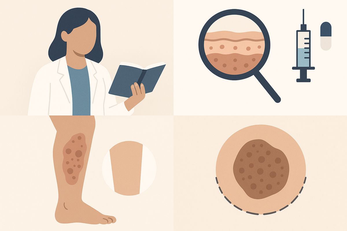
Necrobiosis lipoidica is a rare, chronic inflammatory skin disease. It’s most recognized for causing shiny, reddish-brown patches—often with a yellowish center—primarily on the shins. The full medical term, necrobiosis lipoidica diabeticorum, reflects its strong association with diabetes, but it can occur in people without diabetes as well.
Key Features
- Location: Most commonly affects the lower legs, especially the shins, but can appear elsewhere. Occasionally, lesions develop on the feet, arms, trunk, or even the face, though these locations are much less common.
- Appearance: Lesions begin as small, red-brown papules or bumps that gradually expand into larger, oval, or irregularly shaped patches. Over time, these can become several centimeters in diameter and may merge with nearby lesions, creating larger plaques.
- Texture: Patches are often thin, shiny, and may have a yellowish, waxy center surrounded by a reddish border. The surface is typically smooth, and the skin overlying the patches can appear atrophic (thinned), making underlying veins more visible.
- Other Names: Sometimes called 'shin rash diabetes' or 'brown shin patches' in lay terms. Medical professionals may abbreviate as NL.
Prevalence and Demographics
Necrobiosis lipoidica is uncommon, affecting roughly 0.3% of people with diabetes. It is three times more common in women than men, with most patients diagnosed between ages 20 and 60. Most cases are reported in adults, but children and adolescents can occasionally be affected, especially those with long-standing type 1 diabetes.
This condition does not usually cause pain or itchiness, but complications such as ulceration can develop if the skin becomes fragile or injured. In some studies, up to 35% of cases may eventually ulcerate if not managed. Identifying necrobiosis lipoidica early is key to proper management and preventing further skin breakdown and infection.
Necrobiosis lipoidica is often chronic and persistent. Even with treatment, complete resolution is rare, but early intervention can help control symptoms and reduce the risk of complications. Because of its link with diabetes, it’s also a signal to review overall metabolic control and foot care practices.
Section 2: Recognizing Symptoms and Early Signs
Typical Symptoms
The most common manifestation is the appearance of one or more slowly enlarging patches on the shins. These patches have distinct features:
- Color: Reddish-brown or orange-yellow, sometimes with a paler center. Early lesions may appear more red or pink, becoming more yellowish or brown as they mature.
- Edges: Well-defined, often with a slightly raised, red or purple border. The border may be more pronounced in active or expanding lesions.
- Surface: Smooth, shiny, and thin, sometimes showing visible blood vessels (telangiectasia). The skin may feel cool to the touch and is often fragile.
- Size: Patches can range from less than 1 cm to several centimeters in diameter, expanding over months or years. Lesions may join together to form larger plaques, sometimes covering a significant portion of the shin.
Progression
Early lesions may be mistaken for minor bruises, insect bites, or other rashes. Over time, patches may become more pronounced, with thinning and discoloration. In advanced cases, the skin can break down, leading to open sores or ulcers—especially if bumped or scratched. If an ulcer forms, it may ooze, crust, or become infected, requiring prompt medical attention.
Lesions typically develop slowly. Some people may notice only one or two patches over several years, while others may develop multiple new lesions in a shorter period. The rate of progression can be unpredictable and is not always related to diabetes severity.
Associated Symptoms
Most people do not experience itching or pain, but some report mild discomfort or tenderness, particularly if the lesion is traumatized. In rare cases, there may be a burning sensation or increased sensitivity over the affected area. If ulceration occurs, pain and risk of infection increase significantly.
Other symptoms to watch for:
- Swelling around the lesion, which may indicate inflammation or secondary infection
- Increased sensitivity or burning sensation
- Delayed healing after minor injuries or insect bites
- Development of crusting or oozing if ulcers form
- Thickening or hardening of the skin in chronic lesions
Because necrobiosis lipoidica can resemble other skin conditions, such as eczema, granuloma annulare, or even certain medication-induced rashes, it’s important to monitor any new or changing shin rash carefully. If you notice persistent brown shin patches or shiny, thinning skin, consult a dermatologist or use a digital tool like Rash Detector for initial guidance. However, remember that definitive diagnosis always requires professional evaluation.
Section 3: Necrobiosis Lipoidica and Diabetes Connection
Necrobiosis Lipoidica Diabeticorum Explained
About 60–70% of necrobiosis lipoidica cases occur in people with diabetes, especially type 1 diabetes, but it can also affect those with type 2 or even those without diabetes. The term necrobiosis lipoidica diabeticorum is often used when referring to diabetic patients. While not exclusive to diabetes, the presence of necrobiosis lipoidica can sometimes be the first visible sign of diabetes in some individuals.
Why Does It Happen in Diabetes?
The exact reason is not fully understood, but several factors tie necrobiosis lipoidica to diabetes:
- Changes in small blood vessels (microangiopathy) make the skin more prone to inflammation and poor healing. Diabetes can damage capillaries, reducing blood flow and impairing the skin's ability to repair itself.
- Altered collagen and fat metabolism in diabetes may contribute to the development of lesions. This can result in the breakdown of connective tissue and abnormal lipid (fat) deposits in the skin.
- Immune system changes in diabetes can make skin more susceptible to chronic conditions. Autoimmune reactions may target skin structures, leading to persistent inflammation.
Brown Shin Patches as a Diabetes Warning Sign
While not every person with diabetes will develop necrobiosis lipoidica, its presence can sometimes signal underlying issues with blood sugar control or circulation. Brown shin patches or 'shin rash diabetes' may be an early indicator of diabetic skin changes. If you have diabetes and notice these skin changes, discuss them with your healthcare team to ensure your overall diabetes management is optimized.
For people without a formal diabetes diagnosis, the sudden appearance of necrobiosis lipoidica-like lesions warrants blood sugar screening. Early detection of diabetes can improve long-term outcomes and reduce the risk of complications elsewhere in the body.
For more on how diabetes affects the skin, see our detailed guide on effective skin rash treatments and tools.
In summary, necrobiosis lipoidica serves as both a dermatologic challenge and an important systemic marker for metabolic health. Close collaboration between dermatologists, endocrinologists, and primary care providers is often recommended for best patient outcomes.
Section 4: Causes, Risk Factors, and Pathogenesis
Underlying Causes
While the precise cause of necrobiosis lipoidica remains unclear, research points to several contributory mechanisms:
- Blood Vessel Changes: Damage to small blood vessels (capillaries) in the skin, often related to diabetes, reduces oxygen and nutrient delivery. This can result in chronic low-grade tissue injury and inflammation.
- Collagen Breakdown: Abnormal degradation of collagen and fat in the skin leads to the characteristic lesions. The connective tissue becomes weak, allowing fat to rise towards the surface, giving lesions their yellowish center and atrophic texture.
- Immune Dysregulation: Inflammatory cells accumulate in the skin, perpetuating chronic inflammation and tissue changes. Some studies suggest the involvement of immune complexes and autoantibodies.
Major Risk Factors
- Diabetes Mellitus: The strongest known risk factor, especially long-standing type 1 diabetes. However, individuals with well-controlled diabetes or even those without diabetes can still develop this condition.
- Female Sex: Women are more commonly affected than men, with a reported ratio of about 3:1.
- Age: Most common between ages 20 and 60, but cases in children and older adults have been documented.
- Personal or Family History of Autoimmune Conditions: May increase susceptibility. Conditions such as thyroid disease, rheumatoid arthritis, and lupus have been reported in conjunction with necrobiosis lipoidica.
Other Potential Contributors
- Trauma or repeated injury to the shins, such as from sports, can trigger or worsen lesions due to the Koebner phenomenon (skin trauma leading to new lesions in predisposed individuals)
- Poor circulation or peripheral vascular disease, which may further impair healing and increase risk of ulceration
- Certain medications (though rarely a direct cause), such as corticosteroids or immunosuppressants, which might influence immune function or skin health
- Genetic predisposition, though specific genes have not been clearly identified
Necrobiosis lipoidica is not contagious, nor is it caused by poor hygiene. Understanding your own risk factors can help you monitor for early signs and take preventive steps. For example, people with diabetes—especially those with a history of poor blood sugar control—should inspect their lower legs regularly for new or changing patches.
It’s also important to note that environmental factors, such as prolonged standing, frequent leg trauma, or exposure to harsh weather, may exacerbate symptoms, although these are not direct causes.
Section 5: Diagnostic Tools and When to See a Doctor
How Is Necrobiosis Lipoidica Diagnosed?
Diagnosis is usually based on a combination of medical history, physical examination, and—if needed—skin biopsy. Dermatologists look for the classic appearance and location of lesions. In some cases, additional tests may be performed to rule out similar conditions such as granuloma annulare, sarcoidosis, or cutaneous infections.
- Clinical Exam: Visual inspection and palpation of the lesions. A dermatologist evaluates color, border, texture, and location. They may ask about onset, progression, associated symptoms, and history of trauma or diabetes.
- Skin Biopsy: A small tissue sample is taken to confirm the diagnosis under a microscope. Pathologists look for layers of degenerated collagen, lipid deposits, thickened blood vessels, and chronic inflammatory cells. Biopsy is especially valuable if the appearance is atypical or if the lesion fails to respond to standard therapy.
- Blood Tests: Assess blood sugar levels, especially if diabetes is suspected but not yet diagnosed. Other tests may include HbA1c (for long-term glucose control), lipid profile, and autoimmune screening if other diseases are considered.
In rare cases, imaging studies such as Doppler ultrasound may be performed to assess blood flow if there is concern for underlying vascular disease.
Digital Tools for Rash Identification
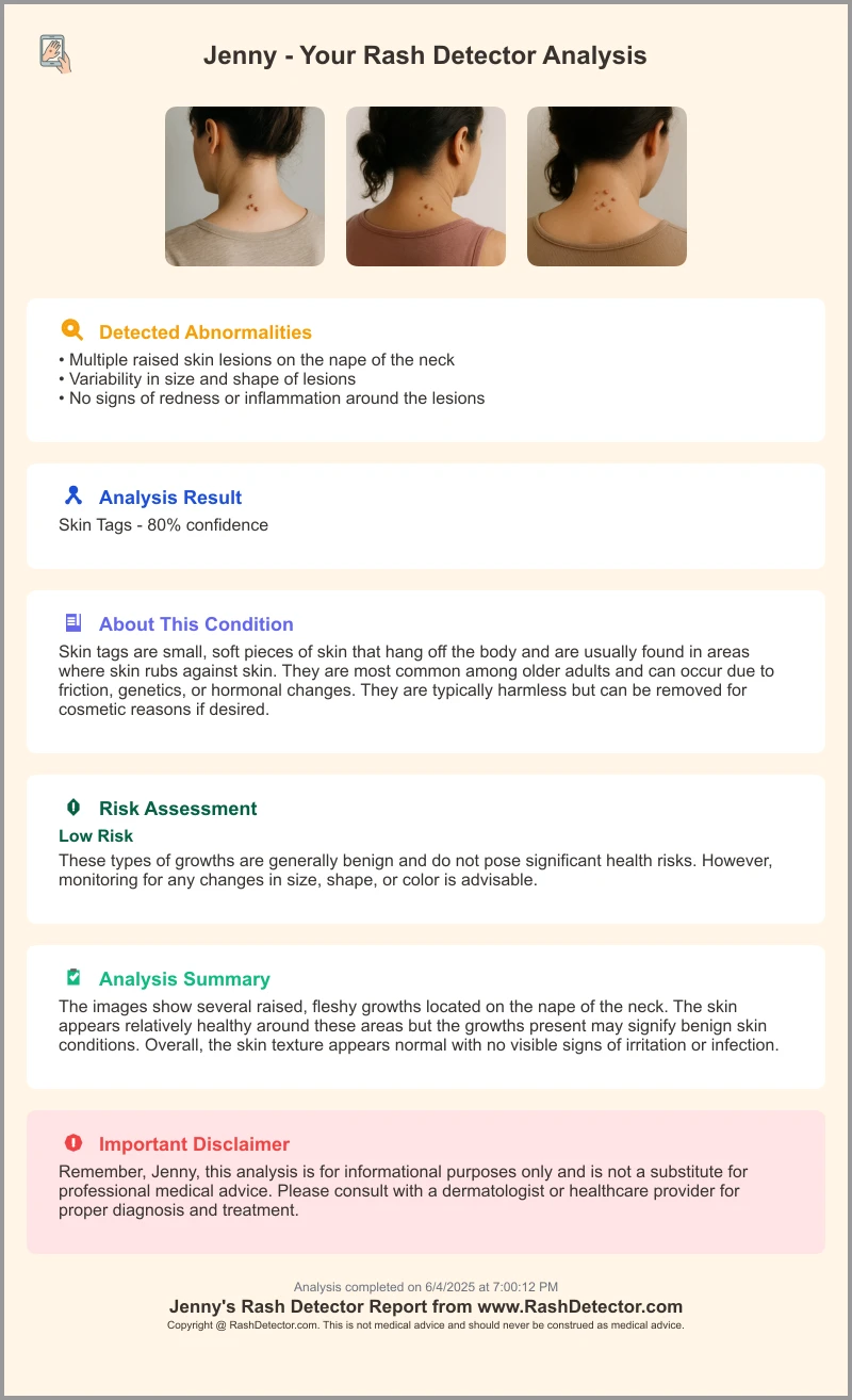
With the rise of AI-powered apps, preliminary rash identification is more accessible. Tools like Rash Detector enable you to upload three images of your rash for instant analysis and guidance. While not a substitute for professional evaluation, these tools can:
- Provide possible causes and initial advice, helping users understand whether their rash may resemble necrobiosis lipoidica or another condition.
- Help track changes over time, allowing users to upload follow-up images and document lesion progression or healing.
- Offer peace of mind or prompt a timely doctor visit if the app suggests a potentially serious or persistent rash.
AI tools can be particularly valuable for people with limited access to dermatologists or those who want guidance before scheduling an in-person appointment. However, they are not designed to replace expert medical evaluation, especially for complex or worsening skin conditions.
When to Seek Medical Care
See a doctor or dermatologist if you notice:
- Patches that change rapidly, ulcerate, or become painful
- Signs of infection (redness, swelling, pus, fever)
- Persistent or spreading lesions that do not improve with gentle skincare
- Skin breakdown, oozing, or non-healing wounds on the legs
- Rashes in unusual locations (such as the face or arms) or with unusual features
Never rely solely on online tools for diagnosis and treatment. Necrobiosis lipoidica can mimic other, sometimes serious, conditions. Early professional evaluation ensures the best outcomes. If lesions are severe, spreading, painful, or ulcerated, seek medical attention immediately.
Section 6: Necrobiosis Lipoidica Treatment and Management
Medical Treatments
There is no universal cure for necrobiosis lipoidica, but several treatments can help control symptoms and prevent complications. Therapies are tailored to the individual and the severity of the condition. Some people may require a combination of treatments or adjustments over time.
- Topical Steroids: First-line treatment to reduce inflammation and slow lesion progression. Applied directly to affected areas, often under occlusion (covered by a dressing) for increased absorption. Commonly used steroids include clobetasol or betamethasone.
- Intralesional Steroid Injections: For thicker or rapidly growing patches, injected steroids can decrease inflammation locally. This is typically performed by a dermatologist in the clinic and may be repeated every few weeks as needed.
- Immunomodulating Creams: Such as tacrolimus or pimecrolimus, sometimes used if steroids are ineffective or not tolerated. These can help reduce inflammation without the side effects of long-term steroid use.
- Oral Medications: In severe or ulcerated cases, oral steroids, antiplatelet drugs (such as aspirin or pentoxifylline), or immunosuppressants (like cyclosporine or methotrexate) may be considered. These are reserved for refractory cases due to potential systemic side effects.
- Wound Care: If ulcers develop, careful wound management and infection prevention are crucial. This may involve regular cleaning, non-adherent dressings, and topical antibiotics if secondary infection is suspected. A wound care specialist may be involved for chronic or large ulcers.
Other Therapeutic Options
- Phototherapy: Ultraviolet light treatments, such as PUVA therapy, may help some patients by reducing inflammation and slowing lesion progression. Phototherapy is generally used in specialized dermatology clinics and requires multiple sessions.
- Surgical Options: Rarely, skin grafting or surgery is required for non-healing ulcers. Surgery is only considered when medical and wound care therapies have failed, and is typically followed by heightened monitoring to prevent recurrence.
- Other Medications: In select cases, medications like nicotinamide, chloroquine, or topical retinoids have been tried with variable success.
Lifestyle and Supportive Care
- Blood Sugar Control: In those with diabetes, good glycemic control is associated with better skin outcomes. Keeping blood glucose levels within target ranges reduces the risk of new lesions and supports healing. Regular monitoring and adjustments to medication, diet, or physical activity may be necessary.
- Protect Skin from Injury: Patches are thin and fragile—avoid bumps and scrapes. Wear long pants, shin guards, or padding during activities that might expose your legs to trauma.
- Moisturizers: Keep skin hydrated to promote healing. Use fragrance-free, hypoallergenic products, and apply them after bathing or washing your legs.
- Smoking Cessation: If you smoke, quitting can improve circulation and skin health, potentially aiding in lesion healing.
- Foot and Leg Inspections: Regularly check for new lesions, ulcers, or signs of infection, especially if you have diabetes or poor sensation in your legs.
Explore more about general skin rash management and over-the-counter options in our guide on the best anti-itch creams.
Note: Always consult your healthcare provider before starting any new treatment. Severe, spreading, or ulcerated lesions require prompt medical attention. Self-treatment is not recommended for active, painful, or infected lesions.
Section 7: Living with Necrobiosis Lipoidica: Daily Care and Prevention
Daily Skin Care
- Gentle Cleansing: Use mild, fragrance-free soaps to avoid irritation. Avoid scrubbing the patches, as this can cause further thinning or trauma.
- Moisturize Regularly: Apply non-greasy, hypoallergenic moisturizers after bathing. Petroleum jelly or specialized barrier creams can be applied to ulcers or at-risk skin to maintain hydration and support healing.
- Sun Protection: Cover affected areas with clothing or sunscreen, as sunburn can worsen lesions. Use broad-spectrum sunscreens with SPF 30 or higher on exposed skin.
- Avoid Irritants: Stay away from perfumes, dyes, and harsh chemicals that could trigger irritation or allergic reactions.
Avoiding Complications
- Do not scratch or pick at patches. The skin is fragile and easily damaged, increasing the risk of ulceration and infection.
- Wear protective gear during sports or activities prone to leg injuries. Shin guards or padded clothing can help prevent trauma.
- Inspect your legs daily for new or changing lesions, especially if you have diabetes. Use a mirror or ask for help if you have difficulty seeing your lower legs.
- Keep your blood sugar levels within recommended targets if you have diabetes.
- Promptly treat any cuts, scrapes, or bug bites to avoid secondary infection.
Emotional and Social Support
Visible skin changes can affect confidence and self-esteem. Consider:
- Seeking support from patient groups or online communities. Organizations for people with diabetes or chronic skin conditions can offer valuable advice and reassurance.
- Discussing concerns with your healthcare provider, who can address both physical and emotional aspects of the condition. Some people benefit from counseling or support groups.
- Exploring cosmetic options (like makeup or skin-tone concealers) if desired, but ensure products are non-irritating and hypoallergenic. Test new products on a small area first.
- Educating friends and family about your condition to reduce stigma and increase understanding.
Necrobiosis lipoidica does not increase your risk for skin cancer or other serious illnesses, but persistent or worsening lesions should always be evaluated. Digital tools like Rash Detector offer a convenient way to monitor your skin, but should never replace medical advice for diagnosis or ongoing management.
With consistent care, most people can manage their symptoms and maintain an active lifestyle. If your condition is affecting your quality of life, ask your doctor about additional resources or referral to a specialist.
Conclusion
Necrobiosis lipoidica is a rare but significant skin condition, particularly for people with diabetes. Recognizing the classic brown shin patches and understanding the link with diabetes enables earlier intervention and better outcomes. While the condition is often persistent, many patients find relief with a combination of medical treatment, good skin care, and careful monitoring.
If you notice new or unusual shin rashes, especially if you have diabetes or risk factors, consult a dermatologist promptly. For initial guidance and to track skin changes over time, try uploading your photos to Rash Detector for instant, AI-powered analysis. Remember, only a medical professional can provide a definitive diagnosis and personalized treatment plan.
Important: For severe, spreading, painful, or ulcerated lesions, seek medical attention immediately.
FAQ
Q: Can necrobiosis lipoidica go away on its own?
A: In some cases, lesions may fade or flatten over time, but most patients experience persistent or recurrent patches. Early treatment can help prevent complications.
Q: Is necrobiosis lipoidica dangerous?
A: The condition itself is not dangerous, but skin breakdown and ulcers can lead to infection. Prompt wound care and medical attention are important if ulcers occur.
Q: What is the difference between necrobiosis lipoidica and other shin rashes?
A: Necrobiosis lipoidica is characterized by shiny, brownish-yellow patches with well-defined borders. Other rashes, such as eczema or allergic reactions, usually look different and may itch. If unsure, consult a dermatologist or use a digital tool for preliminary guidance.
Q: Does controlling my diabetes help with necrobiosis lipoidica?
A: Good blood sugar control can improve overall skin health and may help slow the progression of necrobiosis lipoidica, although it does not guarantee resolution of existing lesions.
Q: Are there any over-the-counter treatments that help?
A: Moisturizers and gentle skincare can help protect affected areas, but prescription medications are often needed for active lesions. For general anti-itch relief, see our recommendations in this anti-itch cream guide. Always check with your healthcare provider before starting new treatments.
衣装を通販で探すときは、商品画像だけでなく実寸とセット内容も確認しておくと安心です。サイズやセット内容を確認する場合は、砂狼シロコ コスプレ衣装から候補を探せます。最後に衣装全体を試着し、正面だけでなく横や後ろからもシルエットを確認すると安心です。