Atypical Skin Rashes Diagnosis: Expert Guide to Unusual Eruptions
Learn how atypical skin rashes diagnosis can reveal underlying conditions and prevent complications. This expert guide explores unusual skin eruptions.
Estimated reading time: 8 minutes
Key Takeaways
- Definition: Atypical rashes deviate from classic patterns in morphology, distribution, or duration.
- Importance: Early recognition can uncover systemic disease or rare conditions and prevent complications.
- Structured Approach: Comprehensive history → Detailed exam → Pattern recognition → Targeted testing.
- Common Pitfalls: Confusing drug eruptions with viral exanthems or overlooking rare presentations.
- Referral: Engage dermatology, rheumatology, or infectious‐disease specialists for complex or refractory cases.
Table of Contents
- Understanding Atypical Skin Rashes Diagnosis
- Diagnostic Approach to Atypical Skin Rashes Diagnosis
- Differential Diagnosis in Atypical Skin Rashes Diagnosis
- Uncommon Presentations and Case Studies in Atypical Skin Rashes Diagnosis
- Treatment and Referral Considerations for Atypical Skin Rashes Diagnosis
- Conclusion and Takeaways on Atypical Skin Rashes Diagnosis
- FAQ
Understanding Atypical Skin Rashes Diagnosis
Atypical eruptions differ from common atopic or viral exanthems by showing unexpected features, persistence, or unusual morphology.
Key differentiators for atypical skin rashes:
- Deviations from classic locations or appearances (e.g., involvement of palms/soles).
- Lesions persisting beyond expected duration (weeks to months).
- Uncommon morphologies (plaque‐like lesions, papules, ichthyosiform changes).
Examples of atypical presentations:
- Adult‐onset Still’s disease: Persistent pruritic plaques instead of fleeting salmon‐pink maculopapular rash.
- Atypical pityriasis rosea: Absence of a herald patch or palm/sole involvement, mimicking other dermatoses.
Sources:
PubMed: Atypical Rash Patterns
PMC Article on Unusual Eruptions
Diagnostic Approach to Atypical Skin Rashes Diagnosis
A systematic, step-wise evaluation helps pinpoint the cause of unusual eruptions.
Step 1: Comprehensive Patient History
- Demographics: age, sex, race.
- Onset and duration: acute vs. chronic.
- Previous episodes and outcomes.
- Medication exposures: prescription, OTC, supplements.
- Travel and environmental exposures.
- Infection history and vaccination records.
- Systemic symptoms: fever, joint pain, weight changes.
Step 2: Detailed Physical Examination
- Color: erythematous, violaceous, hypopigmented.
- Distribution: localized vs. generalized.
- Morphology: macules, papules, plaques, vesicles, pustules.
- Configuration: annular, linear, reticular.
- Mucosal involvement and associated findings (lymphadenopathy, organomegaly).
Step 3: Symptom Assessment and Pattern Recognition
- Constitutional signs suggest systemic disease.
- Pruritus vs. pain vs. burning guides toward allergic, inflammatory, or neuropathic causes.
- Evolution: fixed lesions vs. migrating eruptions.
Step 4: Diagnostic Testing When Uncertain
Ensuring high-quality images enhances AI support; see best photography practices.
- Biopsy and Histopathology: Special stains (PAS, GMS) detect fungi or atypical organisms.
- Laboratory Studies: CBC with differential, ESR/CRP, ANA, rheumatoid factor, viral panels, serum ferritin.
- Imaging: Joint x-rays/MRI, ultrasound for organomegaly.
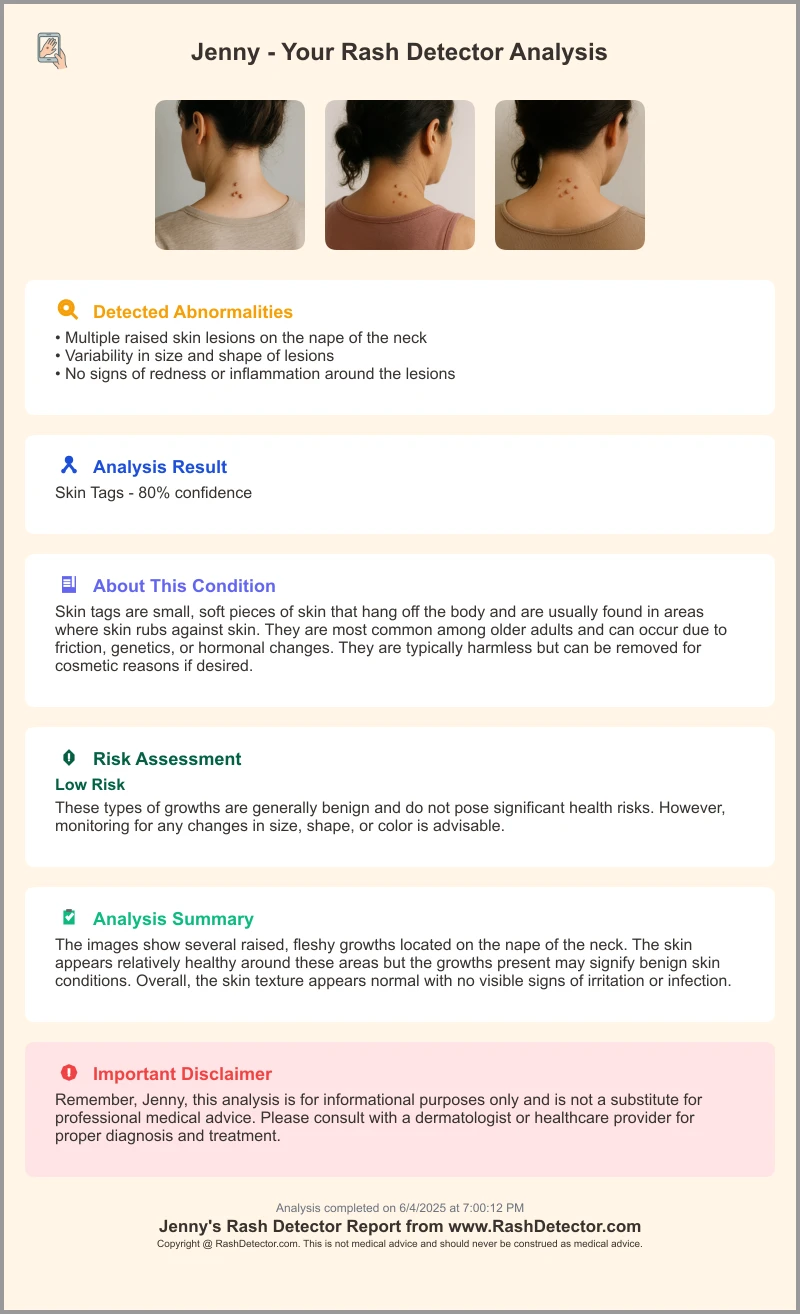
AI analysis can complement traditional tests. By uploading images to tools like Rash Detector, clinicians receive instant assessments:
Sources:
AFP 2010/0315 P726
PMC Atypical Eruptions
PubMed Case Series
Differential Diagnosis in Atypical Skin Rashes Diagnosis
A thorough differential prevents mismanagement and guides correct therapy.
Common pitfalls:
- Drug eruption vs. viral exanthem – both may appear morbilliform.
- Benign dermatosis vs. systemic disease (e.g., fixed drug reaction vs. vasculitis).
- Overlooking rare presentations of common diseases (e.g., lupus panniculitis).
- Missing rare diseases (Sweet syndrome, sarcoidosis).
Systematic exclusion strategy:
- Correlate rash features with timeline and exposures.
- Use targeted labs to rule out infections and autoimmune causes.
- Perform biopsy when clinical features overlap.
- Re-evaluate and refine differential with additional studies.
Sources:
AFP 2010/0315 P726
PubMed Case Series
Uncommon Presentations and Case Studies in Atypical Skin Rashes Diagnosis
Case Study 1: Adult-onset Still’s Disease
- Presentation: High fevers, arthralgia, persistent pruritic plaques.
- Challenge: Misdiagnosed as drug reaction, delayed systemic inflammation recognition.
- Resolution: Elevated ferritin, neutrophilic urticarial dermatitis on biopsy, systemic corticosteroids.
- Learning Point: Use serologic markers and histopathology for confirmation.
Case Study 2: Atypical Pityriasis Rosea
- Presentation: Scaly rash on trunk, palms, and soles without herald patch.
- Workup: Negative RPR/VDRL to exclude secondary syphilis.
- Management: Topical corticosteroids; natural course over 6–8 weeks.
- Learning Point: Perform serologic exclusion when morphology overlaps serious conditions.
Sources:
PMC Atypical Eruptions
PubMed Case Series
Treatment and Referral Considerations for Atypical Skin Rashes Diagnosis
Management is tailored to cause, severity, and systemic involvement.
Treatment Options:
- Topical corticosteroids for localized inflammation.
- Systemic corticosteroids or immunosuppressants for moderate to severe autoimmune eruptions.
- Antihistamines for pruritus relief.
- Targeted biologics (e.g., IL-1 inhibitors in Adult-onset Still’s disease).
- Antimicrobials when infections are identified.
When to Refer:
- Uncertain diagnosis after initial evaluation.
- Suspected systemic involvement.
- Refractory rashes despite adequate therapy.
- Advanced immunological testing, phototherapy, or biologics needed.
Teledermatology can be an option; see the online dermatologist consultation guide.
Conclusion and Takeaways on Atypical Skin Rashes Diagnosis
Atypical skin rashes require vigilance, structured evaluation, and a broad differential. Key points:
- Definition: Eruptions with unusual morphology, distribution, or duration.
- Importance: May reveal systemic disease or rare conditions.
- Approach: History → Exam → Pattern recognition → Targeted testing.
- Pitfalls: Confusing drug eruptions, missing rare presentations.
- Referral: Engage specialists for complex or refractory cases.
Stay current with emerging dermatological research to recognize new atypical patterns.
FAQ
Q: What qualifies a rash as “atypical”?
A: It shows unusual morphology, distribution, or persistence beyond expected timelines compared to classic dermatoses.
Q: When should I consider a skin biopsy?
A: If the lesion’s appearance overlaps multiple conditions or persists despite standard therapy, biopsy and histopathology guide diagnosis.
Q: How do AI tools aid in diagnosis?
A: AI platforms provide instant image-based assessments, complementing clinical evaluation and focusing targeted testing.
Q: When is specialist referral warranted?
A: Refer if diagnosis remains uncertain after initial steps, if systemic involvement is suspected, or if the rash is refractory to treatment.