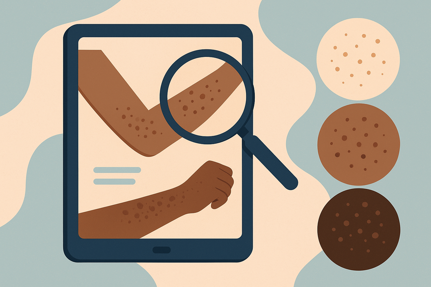
Understanding Skin of Color Rash Characteristics for Accurate Diagnosis
Learn how to recognize skin of color rash characteristics to ensure accurate diagnosis and improved patient care, addressing key differences across diverse tones.

Estimated reading time: 8 minutes
Key Takeaways
- Rashes manifest differently on darker skin, often as purple, gray-brown, or dark patches rather than bright red.
- Post‐inflammatory pigment changes such as hyperpigmentation and hypopigmentation are more pronounced and longer lasting.
- Diagnostic pitfalls arise from under-calling inflammation and lack of training materials featuring skin of color.
- Best practices include dermoscopy, thorough patient history, and texture assessment to improve accuracy.
- Education and increased representation of dark-skin images in curricula are essential for equity.
Table of Contents
- Defining Key Concepts
- Differences Across Skin Tones
- Common Rash Types
- Diagnostic Considerations and Challenges
- Best Practices for Clinicians
- Education and Awareness
- Conclusion
Defining Key Concepts for Skin of Color Rash Characteristics
In order to provide accurate dermatologic care, clinicians must understand what qualifies as “skin of color” and recognize general rash features that may present differently. Skin of color typically refers to medium–dark brown to black skin tones, encompassing individuals of African, Latino, Asian, Middle Eastern, and Indigenous descent, all of which have higher melanin content.
Key rash characteristics include:
- Erythema (redness): Visible as red patches on light skin, but on darker skin it may appear violaceous, ashen gray, or dark brown.
- Inflammation: Swelling, heat, and tenderness that can be underappreciated.
- Scaling: Flaking or peeling of the skin surface.
- Itching: Often leading to scratching and potential secondary infection.
- Texture changes: Roughness, bumps, or thickening (lichenification).
- Post‐inflammatory pigment changes:
- Hyperpigmentation – dark spots persisting after resolution.
- Hypopigmentation – lighter spots from melanocyte damage.
For more examples of how rashes appear on darker skin tones, see this guide on rash appearance in darker skin.
Differences in Skin of Color Rash Characteristics Across Skin Tones
Classic signs such as bright redness may be subtle or absent in skin of color. Clinicians should look for:
- Erythema variability: Light skin shows bright red patches; darker skin may display violaceous, slate-gray, or almost black hues, and whitish spots when perfusion is low.
- Pigmentary changes: Post-inflammatory hyperpigmentation and hypopigmentation last longer and may mask active inflammation.
- Diagnostic challenges: Under-calling inflammation and misreading pigment changes can lead to underestimation of disease severity.
Refer to this practical clinical guide on identifying rashes across diverse skin tones for more details.
Common Types of Skin of Color Rash Characteristics
Top rash types manifest uniquely on darker skin. Visual assessment should emphasize texture, pigment, and distribution:
- Eczema (Atopic Dermatitis): Gray-brown or dark patches with papulation and lichenification rather than redness. Intense itching is common.
- Psoriasis: Plum-colored or dark brown plaques with thick white scaling; edges may be less defined.
- Lichen Planus: Violaceous, flat-topped papules that may blend into brown skin.
- Seborrheic Dermatitis: Brownish or mustard-colored patches on scalp, face, and chest.
- Fungal Infections and Scabies: Gray-brown to black spots in body folds, on feet, or wrists.
Visual tip: Use strong light and dermoscopy to detect subtle vascular patterns and scale.
Diagnostic Considerations and Challenges in Skin of Color Rash Characteristics
Awareness of common errors is key:
- Under-calling inflammation: Less apparent redness may lead to missed severity.
- Pigment confusion: Post-inflammatory hyperpigmentation may be mistaken for active rash.
- Training gaps: Limited textbook images of dark skin perpetuate diagnostic disparities.
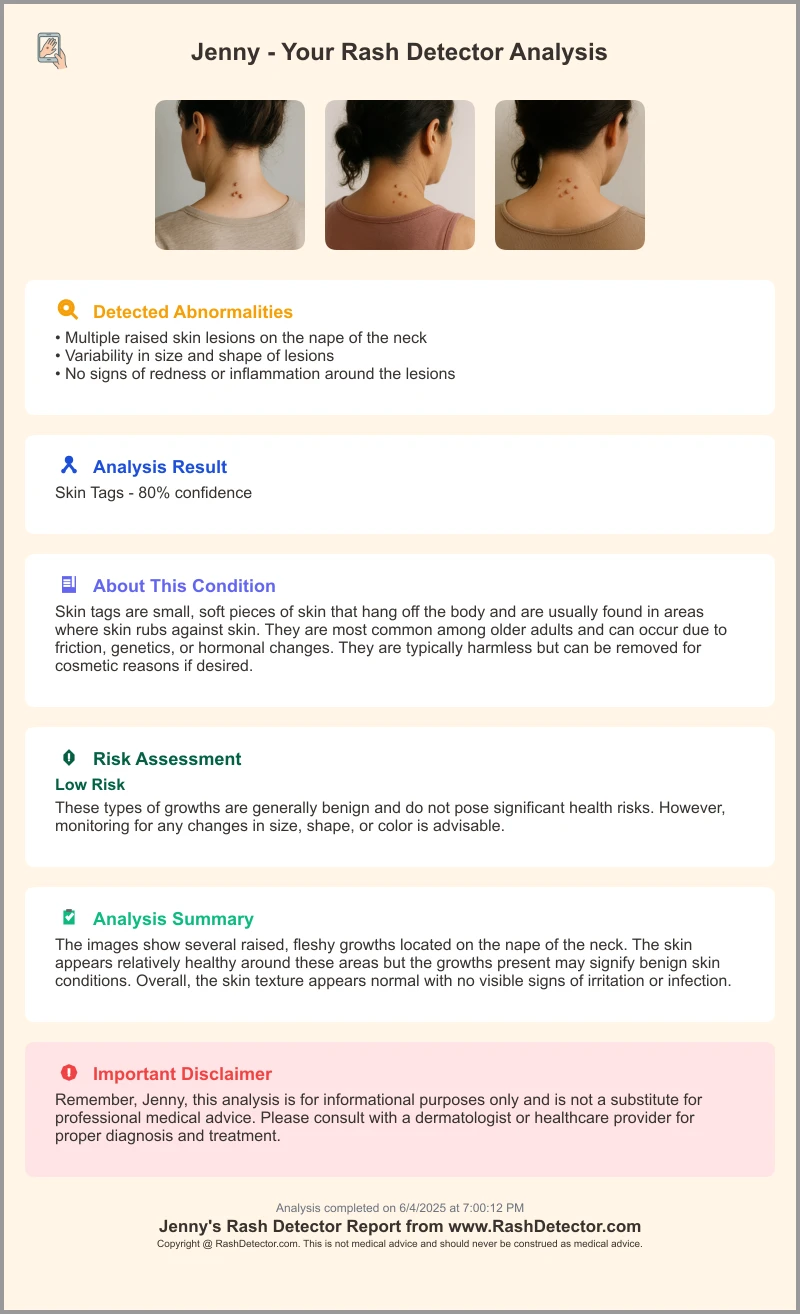
For additional support in visual assessment, clinicians can use AI-assisted tools like Rash Detector to upload clinical images and get instant analysis reports.

Best Practices for Clinicians: Skin of Color Rash Characteristics
Implement these strategies to enhance diagnostic accuracy:
- Holistic assessment: Focus on texture, swelling, and patient-reported symptoms.
- Diagnostic tools: Dermoscopy for subtle vessels and patterns; biopsy for ambiguous lesions; laboratory tests for fungus and scabies.
- Patient history: Document onset, triggers, family history, and separate pigment changes into active vs. post-inflammatory.
- Experience insight: Dermoscopy revealed early psoriasis signs in dark-skinned patients by highlighting microscopic capillaries.
Education and Awareness of Skin of Color Rash Characteristics
Bridging knowledge gaps improves outcomes:
- Training gaps: Most resources focus on light skin, leading to diagnostic oversights.
- Initiatives: The Skin of Color Society offers image libraries and webinars; conferences highlight pigment shifts and dermoscopy dots.
- Call to action: Integrate diverse images into curricula, develop CME modules on dark-skin diagnosis, and encourage case sharing.
Conclusion: Skin of Color Rash Characteristics
Rashes in skin of color often appear purple, gray, or brown rather than red, with significant texture changes and post-inflammatory pigment shifts. By applying informed assessment techniques and advocating for educational equity, clinicians can achieve faster, more accurate diagnoses and reduce health disparities.
FAQ
How does erythema present on darker skin?
On darker skin, erythema may appear violaceous, grayish, or dark brown, and traditional redness can be minimal or absent.
Why are pigment changes important in diagnosing rashes?
Post-inflammatory hyperpigmentation and hypopigmentation often persist after active rash clears, providing clues about prior inflammation and guiding treatment.
What tools aid in evaluating rashes on skin of color?
Dermoscopy, high-intensity lighting, and AI-assisted platforms like Rash Detector can reveal subtle vascular patterns and scale.
How can clinicians improve their skills in this area?
Engage with specialized training from societies like the Skin of Color Society, utilize diverse clinical images, and complete CME modules focusing on skin of color dermatology.


