Rashes in Immunocompromised Patients: Causes, Diagnosis & Management
Explore the causes, diagnosis, and management of rashes in immunocompromised patients, focusing on early recognition and targeted treatment strategies.

Estimated reading time: 8 minutes
Key Takeaways
- Early recognition of rapid spread, atypical morphology, and systemic signs is vital.
- Thorough diagnostics, including cultures, PCR, and biopsy, guide targeted therapy.
- Prompt treatment with appropriate antimicrobials or immunomodulators reduces complications.
- Preventive practices—hygiene, skin checks, and protective measures—minimize risk.
- Timely escalation and specialist involvement improve patient outcomes.
Table of Contents
- I. Introduction to Rashes in Immunocompromised Patients
- II. Understanding Immunocompromise and Skin Vulnerability
- III. Types and Causes of Rashes in Immunocompromised Patients
- A. Infectious Rashes
- B. Non-infectious Rashes
- IV. Recognizing Signs and Symptoms
- V. Diagnostic Approaches
- VI. Treatment and Management Strategies
- VII. Prevention and Best Practices
- VIII. When to Seek Professional Help
- IX. Conclusion: Managing Rashes in Immunocompromised Patients
I. Introduction to Rashes in Immunocompromised Patients
Rashes in immunocompromised patients are skin eruptions or lesions appearing in people whose immune defenses are weakened. A compromised immune system reduces the body’s ability to fight off infections and manage inflammation, making even minor skin changes potentially serious.
Why this matters:
- Unusual infections: Opportunistic bacteria, viruses, fungi, and parasites can cause rashes not seen in healthy people.
- Atypical features: Lesions may lack classic signs (e.g., few vesicles in shingles) or spread rapidly.
- Life-threatening complications: Skin findings can herald sepsis, organ involvement, or systemic spread.
Who is immunocompromised?
- People with HIV/AIDS or other immunodeficiency disorders
- Cancer patients on chemotherapy or radiotherapy
- Recipients of organ transplants on immunosuppressive drugs
- Individuals on long-term corticosteroids or biologic therapies
Early recognition of rashes in immunocompromised patients is critical. Tailored evaluation and prompt treatment can prevent severe outcomes and improve quality of life.
Sources:
https://errolozdalga.com/medicine/pages/FeverRashImmunocomprised.cr.6.3.11.html
https://www.cdc.gov/mpox/hcp/clinical-care/immunocompromised-people.html
II. Understanding Immunocompromise and Skin Vulnerability
Immunocompromise means the body’s natural defenses are impaired. The skin, our largest barrier organ, becomes more vulnerable to infections and injury.
Common causes of immunocompromise:
- HIV/AIDS leading to CD4 cell depletion
- Cancer treatments (chemotherapy, radiotherapy) causing bone marrow suppression
- Autoimmune diseases (lupus, rheumatoid arthritis) treated with immunomodulators
- Organ transplant regimens (calcineurin inhibitors, steroids)
Impact on the skin:
- Lesions may be more severe and deeper (e.g., widespread cellulitis)
- Morphology can be atypical: confluent plaques, necrotic centers, hemorrhagic bullae
- Rapid progression: what starts as a small papule can ulcerate within hours
Increased infection risk:
- Uncommon pathogens (e.g., ecthyma gangrenosum from Pseudomonas)
- Faster systemic spread, leading to higher morbidity and mortality
- Secondary complications like sepsis or multi-organ failure
Understanding how immune suppression alters skin defenses is key to timely diagnosis and effective management of rashes in immunocompromised patients.
Sources:
https://errolozdalga.com/medicine/pages/FeverRashImmunocomprised.cr.6.3.11.html
https://www.cdc.gov/mpox/hcp/clinical-care/immunocompromised-people.html
III. Types and Causes of Rashes in Immunocompromised Patients
Rashes in immunocompromised patients fall into infectious and non-infectious categories. Each has distinct causes and clinical features.
A. Infectious Rashes
- Bacterial infections
- Staphylococcus aureus – folliculitis, abscess formation
- Streptococcus pyogenes – cellulitis, erysipelas
- Pseudomonas aeruginosa – ecthyma gangrenosum: painful red macule → necrotic ulcer
- Viral infections
- Herpes simplex virus (HSV) & herpes zoster virus (VZV): atypical patterns, risk of dissemination
- Mpox: confluent or widespread lesions vs. isolated in immunocompetent hosts
- Cytomegalovirus (CMV): rare cutaneous ulcers, often in transplant recipients
- Fungal infections
- Candida species – intertrigo, diaper‐area satellite pustules
- Dermatophytes – tinea corporis with extensive scaling
- Endemic molds – histoplasmosis or blastomycosis involving skin
- Parasitic infections
- Strongyloides stercoralis – larval tracks in heavy immunosuppression
B. Non-infectious Rashes
- Drug reactions
- Maculopapular eruptions common in polypharmacy
- Severe variants: Stevens-Johnson syndrome (SJS), toxic epidermal necrolysis (TEN)
- Autoimmune or allergic dermatitis
- Lupus dermatitis – photosensitive, malar rash
- Drug-induced lupus-like rash
- Immunocompromised district phenomenon
- Skin areas damaged by prior injury or radiation become new sites for rash development
Recognizing the cause guides targeted therapy. Infections demand antimicrobials, while drug or autoimmune rashes may require immunomodulation.
Sources:
https://errolozdalga.com/medicine/pages/FeverRashImmunocomprised.cr.6.3.11.html
https://www.cdc.gov/mpox/hcp/clinical-care/immunocompromised-people.html
https://pmc.ncbi.nlm.nih.gov/articles/PMC6910813/
IV. Recognizing Signs and Symptoms
Early identification of warning signs can be lifesaving.
Typical presentations:
- Rapidly spreading rash vs. localized lesion
- Atypical morphology:
- Fewer classic vesicles or papules
- Confluent plaques, extensive redness, necrotic or hemorrhagic areas
- Accompanying systemic signs:
- Fever, chills, malaise
- Lymphadenopathy
Red-flag features:
- Painful, necrotic, or hemorrhagic lesions
- High-grade fever or signs of sepsis (tachycardia, hypotension)
- Failure to improve with standard antibiotics or topical treatments
Observation tip: Caregivers should photograph evolving lesions daily to document progression and guide clinicians.
Timely recognition of these signs ensures early intervention for rashes in immunocompromised patients.
Sources:
https://errolozdalga.com/medicine/pages/FeverRashImmunocomprised.cr.6.3.11.html
https://www.cdc.gov/mpox/hcp/clinical-care/immunocompromised-people.html
V. Diagnostic Approaches
A systematic workup improves diagnostic accuracy.
- Medical history
- Onset and progression of the rash
- Current medications, especially immunosuppressants
- Travel history and environmental exposures
- Physical examination
- Lesion morphology: papules, vesicles, ulcers, necrosis
- Distribution: generalized, dermatomal, or localized
- Secondary changes: crusting, scaling, discharge
- Laboratory tests
- Bacterial culture and sensitivity testing
- Viral PCR (HSV, VZV, CMV, mpox) for rapid pathogen ID
- Fungal KOH preparation and culture for yeast or molds
- Skin biopsy
- Indications: progressive, atypical, or severe lesions
- Utility: changes diagnosis in up to 25% of cases, even if treatment remains similar
Early and comprehensive testing shortens time to targeted therapy and reduces complications from rashes in immunocompromised patients.
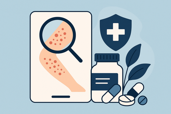
Moreover, AI-assisted solutions such as Rash Detector can expedite analysis by allowing users to upload images and receive an AI-generated report in minutes.
Sources:
https://errolozdalga.com/medicine/pages/FeverRashImmunocomprised.cr.6.3.11.html
https://pubmed.ncbi.nlm.nih.gov/8434974/
VI. Treatment and Management Strategies
Effective care combines targeted therapy, symptom relief, and immune adjustment.
Targeted antimicrobial therapy
- Antibiotics
- Anti-pseudomonal agents (e.g., ceftazidime, ciprofloxacin) for ecthyma gangrenosum
- Anti-staphylococcal regimens (e.g., nafcillin, linezolid) for abscesses
- Antivirals
- Acyclovir or valacyclovir for HSV and VZV
- Tecovirimat for mpox lesions
- Antifungals
- Fluconazole or echinocandins for Candida infections
- Terbinafine for extensive dermatophyte involvement
- Cautious use of steroids or immunomodulators
- For severe drug reactions or autoimmune rashes
- Balance risks: may exacerbate secondary infections
Supportive care
- Analgesics and antipyretics for pain and fever
- Topical wound care:
- Moist dressings, gentle cleansing
- Emollients to maintain skin barrier
Immune management
- Adjust immunosuppressive regimen in consultation with specialists
- Monitor organ function and blood counts
Specialist involvement
- Dermatology for complex biopsy interpretation
- Infectious disease for resistant or unusual pathogens
A multidisciplinary approach ensures comprehensive management of rashes in immunocompromised patients.
Source:
https://errolozdalga.com/medicine/pages/FeverRashImmunocomprised.cr.6.3.11.html
VII. Prevention and Best Practices
Proactive measures reduce risk and severity of skin complications.
Personal hygiene
- Gentle daily cleansing with pH-balanced, fragrance-free soaps
- Pat skin dry; avoid vigorous scrubbing
Environmental precautions
- Avoid contact with individuals who have active infections
- Steer clear of stagnant water, contaminated soil, and animal waste
Protective measures
- Wear gloves when gardening or handling animals
- Use barrier dressings on skin breaks
Skin surveillance
- Routine self-exams: look for new or changing lesions
- Photograph suspicious areas for baseline comparison
Education and reporting
- Teach patients and caregivers early red-flag signs
- Encourage immediate reporting of worsening or persistent rashes
Implementing these best practices can prevent many cases of rashes in immunocompromised patients.
Source:
https://www.cdc.gov/mpox/hcp/clinical-care/immunocompromised-people.html
VIII. When to Seek Professional Help
Knowing red-flag scenarios ensures timely escalation of care.
Immediate medical attention if you observe:
- Rapidly advancing necrosis or deep ulceration
- High-grade fever, chills, hypotension (sepsis indicators)
- Severe pain unrelieved by home analgesics
- Large body-surface involvement or mucosal lesions
- Suspected high-risk pathogens (Pseudomonas, disseminated VZV, mpox)
Guidance for healthcare providers:
- Tailor diagnostic intensity to level of immunosuppression
- Consider inpatient admission for intravenous therapy and close monitoring
- Engage dermatology and infectious disease specialists early
Urgent evaluation and management can significantly improve outcomes for rashes in immunocompromised patients.
Sources:
https://errolozdalga.com/medicine/pages/FeverRashImmunocomprised.cr.6.3.11.html
https://www.cdc.gov/mpox/hcp/clinical-care/immunocompromised-people.html
IX. Conclusion: Managing Rashes in Immunocompromised Patients
Early recognition, accurate diagnosis, and targeted treatment are key to minimizing complications. A proactive partnership between patients, caregivers, and healthcare teams supports better outcomes.
- Recognize rapid spread, atypical morphology, and systemic signs
- Employ thorough diagnostics: cultures, PCR, biopsy
- Start targeted antimicrobials or immunomodulatory therapy promptly
- Practice preventive hygiene and routine skin checks
- Know when to escalate care and involve specialists
Additional Resources:
CDC immunocompromised care guidelines
PubMed review on skin biopsy utility
FAQ
- What causes rashes in immunocompromised patients?
A weakened immune system—whether from HIV, cancer therapy, transplants, or immunosuppressive drugs—allows opportunistic pathogens and atypical inflammatory processes to affect the skin. - How are these rashes diagnosed?
Through a combination of detailed history, physical exam, laboratory tests (cultures, PCR, KOH), and often a skin biopsy to confirm the underlying cause. - What treatments are recommended?
Targeted antimicrobial therapy (antibiotics, antivirals, antifungals), cautious immunomodulation for drug reactions or autoimmune rashes, plus supportive wound care and symptom relief. - How can I prevent these skin complications?
Maintain gentle hygiene, avoid exposure to active infections or contaminated environments, perform routine skin checks, and educate caregivers on early warning signs. - When should I seek professional help?
If you notice rapidly progressing lesions, high-grade fever, signs of sepsis, severe pain, or extensive skin involvement—especially with necrosis or hemorrhage.