Necrobiosis Lipoidica Diabeticorum Treatment: A Comprehensive Guide
Discover effective necrobiosis lipoidica diabeticorum treatment options to manage this chronic skin condition in diabetic patients, improving quality of life.
Estimated reading time: 8 minutes
Key Takeaways
- Necrobiosis lipoidica diabeticorum (NLD) is a chronic inflammatory skin disorder linked to diabetes, often affecting the shins.
- Early recognition and a tailored treatment plan can prevent ulceration and secondary infections.
- Treatment options range from topical corticosteroids and immunomodulators to systemic agents, laser therapy, and surgical interventions.
- Glycemic control and healthy lifestyle modifications support healing and may slow lesion progression.
- A multidisciplinary approach—dermatologist, endocrinologist, wound care—optimizes outcomes.
Table of Contents
- Introduction
- Understanding the Condition
- Exploring Treatment Options
- Role of Glycemic Control
- Integrating Patient-Specific Considerations
- Evidence-Based Approach
- Practical Guidance and Lifestyle Modifications
- Conclusion
- FAQ
Introduction
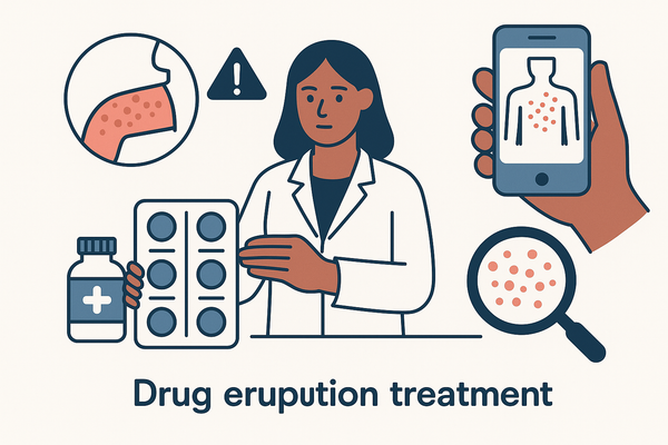
Necrobiosis lipoidica diabeticorum treatment focuses on managing a persistent skin disorder in people with diabetes. NLD often appears as well-defined, shiny red-brown or yellowish patches that can ulcerate if left untreated. A clear care plan helps reduce complications and improve quality of life.
Understanding the Condition
Effective management begins with recognizing the pathology and presentation:
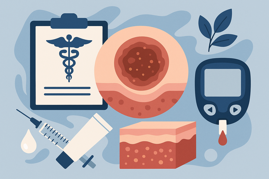
Definition and Pathology
- Necrobiosis refers to collagen degeneration in the dermis.
- Granulomatous inflammation surrounds necrotic collagen fibers.
- Thickened vessel walls impair microcirculation, exacerbating lesions.
- Learn more in the WWIC understanding NLD.
Characteristic Lesions
- Well-demarcated plaques with an atrophic yellow center and red-violet border.
- Begin as firm papules that coalesce and flatten.
- Telangiectasia often visible over the center.
- See the DermNet NZ overview.
Common Symptoms and Complications
- Often asymptomatic initially; may develop itching or tenderness.
- Minor trauma can lead to ulceration and secondary infection.
- Cosmetic concerns may impact self-esteem.
- Higher prevalence in women and smokers.
- Additional details at the MedlinePlus article.
Exploring Treatment Options
No universal protocol exists; therapy is tailored by lesion age, depth, and patient factors.
Topical Therapies
High-Potency Topical Corticosteroids
- Mechanism: Block inflammatory cytokines via glucocorticoid receptors.
- Application: Thin layer once or twice daily at lesion edges.
- Response: Often 4–8 weeks to visible improvement.
- Side Effects: Skin atrophy, telangiectasia, striae.
- Reference: Medical News Today.
Topical Immunomodulators
- Tacrolimus 0.1% ointment reduces T-cell activation.
- Use twice daily when steroids are contraindicated or ineffective.
- Monitor for local burning sensation.
- Details at the DermNet NZ overview.
Systemic Therapies
- Oral Corticosteroids: Prednisone 0.5–1 mg/kg/day, taper slowly; watch for osteoporosis and hyperglycemia. See the WWIC guide.
- Antiplatelet Agents: Aspirin (75–150 mg daily) or pentoxifylline (400 mg TID) to enhance microcirculation.
- Immunosuppressants/Biologics: Cyclosporin (2–5 mg/kg/day) or anti-TNF agents for refractory cases.
Advanced/Procedural Interventions
- Intralesional Corticosteroid Injections: Triamcinolone 5–10 mg/mL every 4–6 weeks.
- Laser Therapy: Pulsed Dye Laser (585 nm, 7–10 J/cm²; 4–6 sessions).
- Phototherapy: UVA1 or nb-UVB over 8–12 weeks.
- Surgical Excision and Skin Grafting: For non-healing ulcers or thick plaques.
Role of Glycemic Control
Tight blood sugar management (HbA1c < 7%) supports lesion healing and may slow progression. For tips on diabetic skin rash management, see our guide.
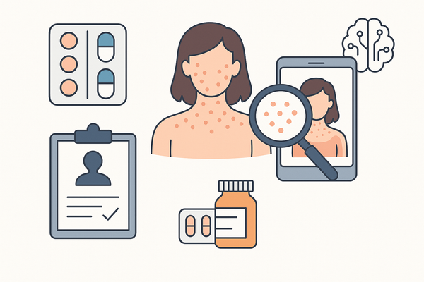
Many patients use AI tools like Rash Detector for photo tracking and instant reports.
Integrating Patient-Specific Considerations
A personalized plan accounts for:
- Lesion size, depth, and ulceration status.
- Comorbidities such as vascular and renal disease.
- Previous treatment responses and tolerance.
- Patient lifestyle, occupation, and preferences.
Stepwise Algorithm
• Early non-ulcerated lesions: high-potency topicals ± tacrolimus.
• Moderate/severe or steroid-resistant: systemic agents (aspirin, pentoxifylline, low-dose steroids).
• Ulcerated/chronic lesions: intralesional therapy, laser/phototherapy, surgical referral.
Multidisciplinary Coordination
Dermatologist, endocrinologist, and wound care nurse collaboration is essential.
Evidence-Based Approach
Research is limited to small studies and case series:
- High-potency steroids: X% reduction in redness at 8 weeks.
- Pentoxifylline: Y% improvement in ulcer healing over 12 weeks.
- Pulsed Dye Laser: 50% cosmetic score improvement after 4 sessions.
Emerging biologics and advanced lasers show promise but need larger trials.
Practical Guidance and Lifestyle Modifications
Skin Care Routine
- Use mild, fragrance-free cleansers.
- Apply emollients twice daily to support barrier function.
- Avoid scratching or trauma to lesions.
Wound Management for Ulcers
- Gentle debridement of non-viable tissue.
- Non-adherent, moisture-balanced dressings.
- Monitor for signs of infection.
Dietary Advice
- Favor low glycemic index foods: whole grains, vegetables.
- Balance macros: lean protein, healthy fats, complex carbs.
- Consult ADA meal-planning resources for portions.
Exercise Recommendations
- 150 minutes/week of moderate aerobic activity.
- Improves circulation and glucose control.
- Include lower-limb stretching to minimize trauma risk.
Smoking Cessation
- Smoking impairs wound healing and worsens lesions.
- Benefits begin within weeks of quitting.
- Refer to local support programs or NHS Quit Smoking guidelines.
Conclusion
Necrobiosis lipoidica diabeticorum treatment requires a patient-tailored, multimodal strategy:
- Combine topical, systemic, and procedural therapies to address inflammation, vascular changes, and immune factors.
- Tight glycemic control and healthy habits support recovery.
- Regular follow-up with a multidisciplinary team ensures optimal care.
Consult your dermatologist or endocrinologist to develop an individualized NLD treatment plan.
FAQ
- What causes necrobiosis lipoidica diabeticorum?
The exact cause is unknown, but microvascular changes and immune-mediated collagen degeneration play key roles. - How is NLD diagnosed?
Diagnosis is clinical, supported by biopsy showing granulomatous inflammation and collagen necrobiosis. - Can NLD be prevented?
While prevention is not guaranteed, good glycemic control and avoiding trauma to the shins may reduce risk.