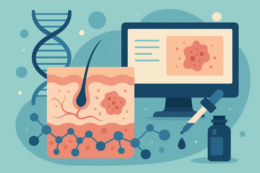
Molecular Markers for Rash Diagnosis: Revolutionizing Precision in Dermatology
Explore how molecular markers for rash diagnosis are transforming dermatology, enhancing precision, and enabling personalized treatments for complex skin conditions.
Estimated reading time: 10 minutes
Key Takeaways
- Molecular markers provide objective, quantitative data for more accurate rash diagnosis.
- Categories include genetic, protein/metabolic, cytokine, microRNA and pathogen‐specific markers.
- Integrating molecular panels with traditional exams enhances sensitivity and specificity (>95%).
- Robust laboratory workflows—PCR, IHC, sequencing—ensure rapid, actionable results.
- Clinical studies demonstrate early detection of CTCL, vitiligo severity assessment and rapid fungal identification.
- Key challenges: cost, technical expertise, validation gaps and standardization across labs.
- Future trends: multi‐omics integration, AI‐driven diagnostics and point‐of‐care molecular tests.
Table of Contents
- Introduction
- Section 1: What Are Molecular Markers?
- Section 2: Understanding Rash Diagnosis
- Section 3: Traditional vs. Molecular Diagnostic Approaches
- Section 4: Key Molecular Markers in Rash Diagnosis
- Section 5: Diagnostic Techniques & Workflow
- Section 6: Research & Clinical Evidence
- Section 7: Challenges & Limitations
- Section 8: Future Perspectives
- Conclusion
- FAQ
Introduction
Molecular markers for rash diagnosis are transforming how clinicians identify and treat skin eruptions. Accurate rash diagnosis matters because rashes can signal serious systemic diseases—from autoimmune disorders to cutaneous cancers. Relying solely on appearance and biopsy can lead to misdiagnosis or delays. Molecular markers offer greater diagnostic precision compared to traditional methods, delivering objective, quantitative data that guide early interventions (PMC 11049353).
In this post, we will:
- Define molecular markers and their diagnostic role
- Review clinical features and hurdles in rash evaluation
- Compare conventional exams with molecular approaches
- Detail gene panels, proteins, cytokines, microRNAs and pathogen‐specific markers
- Explain laboratory workflows: PCR, immunohistochemistry, sequencing
- Highlight key clinical studies and evidence
- Discuss cost, validation gaps and technical challenges
- Explore future trends in personalized dermatology and AI‐driven diagnostics
Section 1: What Are Molecular Markers?
Molecular markers are identifiable DNA, RNA, protein or metabolic features that correlate with a specific biological state or disease. They serve as precise flags in medical diagnostics, offering objective readouts beyond the eye and microscope.
Role in Medical Diagnostics
- Objectivity: Numeric or binary outputs reduce subjective interpretation.
- Quantitative Data: Marker levels can track disease burden and prognosis.
- Early Detection: Subclinical changes are detectable before full symptoms emerge.
Categories of Molecular Markers
- Genetic markers
- Gene mutations (e.g., single nucleotide variants in oncogenes)
- Gene expression panels (mRNA arrays that classify disease subtypes)
- Protein/metabolic markers
- Cytokines (immune signaling proteins like IL-6, TNF-α)
- Metabolites (small‐molecule byproducts indicating cellular stress)
By integrating molecular markers for rash diagnosis, dermatologists gain a robust toolkit for precision medicine, combining genomic, transcriptomic and proteomic insights.
Section 2: Understanding Rash Diagnosis
A “rash” is any visible change in skin—redness, bumps, scaling or blisters. Key clinical features include:
- Eruption pattern (localized vs. diffuse)
- Color and morphology (macules, papules, vesicles)
- Scaling, crusting or lichenification
- Pruritus (itchiness) and discomfort
- Distribution (flexural, acral, mucosal)
Common Rash Etiologies
- Inflammatory
- Psoriasis (silver scales on extensor surfaces)
- Atopic dermatitis (eczema with excoriations)
- Allergic
- Contact dermatitis (poison ivy, nickel allergy)
- Infectious
- Viral exanthems (measles, rubella)
- Fungal infections (tinea corporis)
- Neoplastic
- Cutaneous T-cell lymphoma (CTCL with patches and plaques)
Diagnostic Challenges
Overlapping appearances—psoriasis vs. chronic dermatitis—can confuse clinicians. Early lesions lack hallmark features. Histopathology may also yield inconclusive or borderline findings. By introducing molecular markers for rash diagnosis, we can clarify ambiguous cases and accelerate targeted treatment (PMC 11049353).
Section 3: Traditional vs. Molecular Diagnostic Approaches
Traditional Methods
- Clinical Examination
- Strengths: Immediate, low cost, non‐invasive
- Limitations: Subjective; experience‐dependent
- Histopathology
- Strengths: Tissue architecture, cellular detail
- Limitations: Sampling error; inter‐observer variability
Molecular Approaches
- Higher sensitivity/specificity for specific markers
- Detect subclinical disease (e.g., minimal residual CTCL)
- Quantitative thresholds guide staging and prognosis
- Early identification of pathogenic agents (viral/fungal PCR panels)
Conceptual Comparison Table:
Method | Sample Type | Turnaround | Accuracy | Limitations
Clinical Exam | Visual | Minutes | 60–70% | Subjective
Histopathology | Biopsy | 1–2 days | ~80% | Sampling error
Molecular Panel | Biopsy/Blood | Hours–Days | >95% | Cost, expertise
For further reading on hybrid diagnostic models, see this overview.
To streamline both traditional and molecular diagnostics, clinicians are turning to mobile AI tools like Rash Detector, which analyze rash images in seconds to provide instant preliminary insights alongside lab data.

By leveraging molecular markers for rash diagnosis alongside clinical data, dermatologists can achieve unmatched diagnostic precision and personalized care.
Section 4: Key Molecular Markers in Rash Diagnosis
A. Gene Panels for Cutaneous T-Cell Lymphoma (CTCL)
Researchers have identified an 8–17 gene set differentially expressed in CTCL vs. benign dermatoses, including IL2RA, CCR4, STAT5A, TOX and PLS3. Panels achieve up to 100% sensitivity and >95% specificity when measured by qPCR or RNA sequencing.
B. Protein Markers in Inflammatory Dermatoses
Quantifiable proteins such as S100B, S100A9 and HMGB1 are assayed by ELISA or IHC to stage severity and map inflammatory hotspots.
C. Cytokines and microRNAs
Key cytokines (IL-6, TNF-α) and microRNAs (miR-423, miR-182) correlate with disease activity and can be quantified in serum, plasma or biopsy RNA.
D. Pathogen‐Specific Molecular Markers
PCR targets fungal ribosomal DNA (e.g., Coccidioides spp.) and multiplex viral panels (measles, rubella, parvovirus B19) outperform culture in sensitivity and speed.
Section 5: Diagnostic Techniques & Workflow
Sample Collection & Preparation
- Skin Biopsy: Punch/excisional biopsy; snap-freeze or RNAlater; avoid overfixation.
- Blood Draw: Serum or EDTA plasma; centrifuge and store at –80 °C for cytokine/microRNA assays.
Detection Methods
- PCR/qPCR: Extraction, primer design, amplification and Ct-based quantification.
- Immunohistochemistry (IHC): Antigen retrieval, antibody staining, intensity scoring (0–3+).
- Sequencing: Microarrays or NGS with bioinformatics pipelines for differential expression.
Data Interpretation & Reporting
Standardized reports include numeric fold changes, clinical significance, differential diagnosis and therapeutic recommendations.
Section 6: Research & Clinical Evidence
Case Study A: CTCL Gene Panel Validation
- 100 skin lesions (50 CTCL, 50 benign); 8‐gene qPCR panel; sensitivity 98%, specificity 95%.
- Detected early‐stage CTCL pre‐histologic changes (p < 0.001).
Case Study B: Vitiligo Marker Correlation
- 60 patients; S100B/S100A9 levels correlated with BSA involvement (r=0.82, p < 0.0001).
- ELISA cutoffs guide phototherapy dosing.
Case Study C: Rapid Fungal Detection
- Coccidioides spp.: PCR in 6 hours (92% sensitivity) vs. culture in 10–14 days (65%).
Section 7: Challenges & Limitations
Cost & Accessibility
- Per‐test cost: $100–$500 for panels or multiplex PCR.
- Insurance coverage: variable; prior authorizations often required.
- Resource‐limited settings: lack of NGS/qPCR infrastructure.
Technical Expertise & QC
- Trained molecular pathologists and technologists.
- Accreditation: CLIA/CAP standards; proficiency testing.
Validation Gaps
- Marker variability: limited large‐scale validation.
- False positives/negatives: overlapping expression profiles.
- Standardization: protocol and cutoff discrepancies.
Section 8: Future Perspectives
Personalized Dermatology
Molecular “fingerprint” libraries and tailored therapies matched to individual profiles will drive next‐generation care.
Emerging Technologies
Multi‐omics integration and AI algorithms will unlock complex data patterns for diagnostic insights (PMC 11049353).
Integrated Diagnostics
Digital dermoscopy overlaid with molecular readouts and real‐time point‐of‐care assays will enable comprehensive workflows.
Conclusion
Molecular markers for rash diagnosis are revolutionizing the detection, classification and treatment of cutaneous diseases. By moving beyond subjective exams and biopsy, these biomarkers enhance early detection, improve diagnostic precision and support personalized care. Integrating gene panels, protein assays, cytokine profiles and pathogen‐specific tests into routine workflows shortens time to diagnosis, reduces misclassification and optimizes patient outcomes. We encourage dermatologists, pathologists and laboratory directors to adopt validated molecular marker panels as part of a precision medicine strategy. Embrace molecular diagnostics today to deliver safer, faster and more personalized care for complex or atypical rashes.
FAQ
-
What are molecular markers and why are they important in rash diagnosis?
Molecular markers are measurable DNA, RNA or protein features that correlate with disease states. They offer objective, quantitative data that improve diagnostic accuracy and enable early detection.
-
How do molecular approaches compare to traditional methods?
While clinical exams and histopathology remain valuable, molecular panels achieve higher sensitivity/specificity (>95%), detect subclinical disease and guide prognosis with quantitative thresholds.
-
What are the main challenges in adopting molecular diagnostics?
Key barriers include cost, the need for specialized infrastructure and expertise, variable insurance coverage and a lack of standardized protocols across laboratories.
-
How will AI and multi-omics shape the future of dermatology?
Machine learning will integrate genomics, proteomics and imaging data to uncover complex patterns, enabling real-time, personalized diagnostics and guiding targeted therapies.