Innovations in Rash Detection: Transforming Dermatology with Emerging Technologies
Explore how innovations in rash detection, including AI and imaging, are revolutionizing dermatology to enhance diagnosis and patient outcomes.

Estimated reading time: 8 minutes
Key Takeaways
- Advanced imaging and AI-driven analytics are enhancing the accuracy and speed of rash diagnosis.
- Mobile health integration allows remote monitoring and teledermatology consultations.
- Emerging tools like digital dermatoscopes and OCT deliver non-invasive, high-resolution insights.
- Clinical adoption faces regulatory, privacy, and ethical challenges, but continued research promises standardized solutions.
Table of Contents
- Section 1: Background on Rash Detection
- Section 2: Overview of Current Innovations
- Section 3: Integration with Mobile Health
- Section 4: Emerging Technologies in Detail
- Section 5: Real-World Applications and Case Studies
- Section 6: Challenges and Considerations
- Section 7: Future Outlook and Trends
- Conclusion
- FAQ
Section 1: Background on Rash Detection
Keyword in header: innovations in rash detection
Traditional methods for rash detection rely on:
- Visual inspection by dermatologists
- Patient history review and symptom discussion
- Physical examination under clinical lighting
- Skin biopsy in uncertain or severe cases
Limitations of conventional techniques include:
- Subjectivity and inter-observer variability among clinicians
- Difficulty distinguishing visually similar conditions (e.g., eczema vs. psoriasis)
- Invasiveness, discomfort, and risk of infection from biopsies
- Potential for missed or delayed diagnoses in early or mild presentations
These constraints create a diagnostic gap that innovations in rash detection aim to fill by offering objective, non-invasive, and reproducible tools for clinicians and patients alike.
Section 2: Overview of Current Innovations
Keyword in header: innovations in rash detection
Subsection A: Diagnostic Imaging Advances
- Total-Body Digital Photography (TBDP) + Dermoscopy
– High-resolution, full-body imaging captures lesion changes over time.
– Portable dermatoscopes attach to smartphones for image capture and AI-driven lesion analysis.
– Instant upload to cloud platforms enables remote expert review.
Source: top dermatology technology trends for 2025 - Fluorescence Imaging & Confocal Microscopy
– Fluorescence dyes highlight microstructures and cell activity in skin layers.
– Confocal microscopy provides cellular-level imaging in real time without biopsies.
Source: top dermatology technology trends for 2025 - Optical Coherence Tomography (OCT)
– Non-invasive, depth-resolved imaging offers cross-sectional views of epidermis and dermis.
– Useful for monitoring lesion depth and vascular patterns in inflammatory and neoplastic rashes.
Source: top dermatology technology trends for 2025
Subsection B: AI & Machine Learning
Modern AI models combine convolutional neural networks (CNNs) with patient metadata to classify rashes. Deep learning algorithms analyze thousands of dermoscopic images, detecting subtle patterns beyond human vision. Studies show AI can match or exceed dermatologist accuracy in identifying conditions like melanoma, eczema, and psoriasis. Automated diagnostic suggestions appear within seconds, streamlining workflow and reducing appointment durations (for a detailed overview).
Section 3: Integration with Mobile Health
Keyword in header: innovations in rash detection
- Automated Image Capture
– Guided photo protocols help users take standardized, high-quality rash images using smartphones. - AI Analysis
– Cloud-based algorithms provide instant feedback on rash severity and possible diagnoses. - Teledermatology Video Calls
– Real-time video assessments connect patients with dermatologists anywhere.
Benefits of mHealth integration:
- Remote monitoring reduces in-office visits and travel burdens.
- Access to specialist advice in underserved or rural areas.
- Continuous patient engagement with reminders for photo submissions and follow-up.
- Potential to flag urgent cases for expedited care.
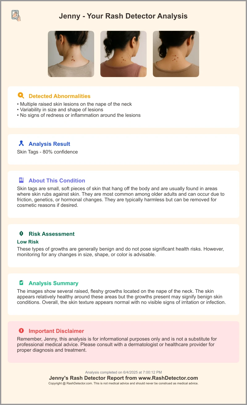
For example, the Skin Rash App leverages AI to analyze user-submitted images and generate comprehensive reports in seconds. Below is a sample report highlighting key diagnostic metrics and recommendations:
Section 4: Emerging Technologies in Detail
Keyword in header: innovations in rash detection
Subsection A: Digital Dermatoscopes
- Device Description
– Handheld, high-magnification camera with polarized light source.
– Built-in cloud connectivity for image upload and AI assessment. - Functionality
– Instant lesion analysis via embedded deep learning models.
– Automated measurement of lesion dimensions and color metrics. - Real-World Usage
– Deployed in primary care settings to triage suspicious lesions before specialist referral. - Source: top dermatology technology trends for 2025
Subsection B: High-Resolution Imaging Systems
- Confocal Microscopy
– Non-invasive, sub-cellular visualization of skin architecture in vivo.
– Resolution: ~1 μm lateral, enabling detection of cellular anomalies.
– Use Cases: Early detection of basal cell carcinoma, mapping inflammatory cell infiltration. - Optical Coherence Tomography (OCT)
– Depth-resolved scans up to 2 mm beneath skin surface without contrast agents.
– Resolution: ~10–15 μm axial, ideal for assessing lesion thickness and vascular patterns.
– Diagnostic Uses: Psoriasis plaque thickness, early melanoma staging. - Source: top dermatology technology trends for 2025
Subsection C: Algorithm-Based Assessments
Neural networks trained on tens of thousands of labeled rash images differentiate conditions such as psoriasis, eczema, fungal infections, and viral exanthems. Performance metrics include up to 92% sensitivity for fungal infections and 95% specificity for melanoma detection, outperforming average clinician rates. These algorithms are embedded in dermatoscopes and mHealth platforms for point-of-care decision support (see technical details).
Section 5: Real-World Applications and Case Studies
Keyword in header: innovations in rash detection
Case Study 1: Onychomycosis Detection via AI Model
Challenge: Nail fungal infections often misdiagnosed or require lengthy culture tests. AI solution: CNN trained on 15,000 nail images. Outcome: Sensitivity improved from 70% to 90%; specificity from 75% to 93%.
Source: review published in Dermatology Times
Case Study 2: Bacterial Skin Disease Identification
Conditions: Leprosy, acne, impetigo. AI Approach: Ensemble model analyzing rash morphology and distribution. Impact: Early leprosy detection reduced delays by 40%; improved acne variant diagnosis.
Source: review published in Dermatology Times
Case Study 3: Public Health Surveillance—EPIWATCH for Monkeypox
Workflow: Smartphone image capture → AI triage → Automated public health alerts. Results: Early identification enabled rapid isolation and contact tracing, decreasing outbreak growth by 25% in pilot regions.
Source: review published in Dermatology Times
Section 6: Challenges and Considerations
Keyword in header: innovations in rash detection
- Regulatory Hurdles
– FDA/CE Approval: AI algorithms and imaging devices must demonstrate safety and efficacy through trials and post-market surveillance.
Source: top dermatology technology trends for 2025 - Data Privacy & Security
– HIPAA/GDPR Compliance: Secure storage, encryption, access controls, anonymization.
– Patient Consent: Transparent policies and opt-in consent for data use. - Clinical Integration
– Training and Adoption: Clinician education on device use and AI interpretation.
– Workflow: EHR integration to ensure tools augment clinical judgment. - Ethical Balance
– Explainable AI to clarify decision factors.
– Bias monitoring across diverse populations to ensure equitable care.
Section 7: Future Outlook and Trends
Keyword in header: innovations in rash detection
- AI Advancement Trajectory
– Deeper AI–EHR integration for automated rash flagging.
– Point-of-care devices offering instant bedside diagnostics. - Wearable & Remote Monitoring
– Smart patches with microfluidic sensors for continuous biomarker tracking.
– Connected smartbands with thermal/optical sensors for early rash detection. - Cross-disciplinary Collaboration
– Dermatologists, engineers, and regulators creating standardized validation frameworks and responsible AI policies.
Stay informed on emerging dermatology tech to harness these innovations in rash detection for better patient outcomes and healthcare efficiency.
Conclusion
Innovations in rash detection are overcoming the limitations of traditional visual exams and biopsies by leveraging AI, machine learning, high-resolution imaging, and mobile health. While regulatory, privacy, and ethical challenges remain, ongoing research and collaboration promise a future where early, precise rash detection becomes standard practice.
FAQ
- How accurate are AI-based rash detection tools?
Studies report AI matching or exceeding dermatologist accuracy, with sensitivity up to 95% for melanoma and 92% for fungal infections. - What privacy safeguards are in place for patient images?
Platforms comply with HIPAA/GDPR, employing encryption, secure storage, and user consent protocols. - When can clinicians expect widespread adoption?
Adoption depends on regulatory approvals and clinical integration efforts, but pilot programs suggest broader use within 2–3 years.