Assessing the Real World Effectiveness of AI Rash Detection in Clinical Practice
Explore the real world effectiveness of AI rash detection and its impact on clinical practice, patient care, and dermatological diagnosis efficiency.
Estimated reading time: 8 minutes
Key Takeaways
- AI rash detection leverages machine learning, CNNs, and computer vision for rapid and standardized skin assessments.
- Real-world studies show up to 92% accuracy and throughput of 500 images/hour, matching or exceeding dermatologists.
- Challenges remain around skin tone biases, data quality, and regulatory compliance, requiring diverse datasets and transparent validation.
- Teledermatology and federated learning are key future directions for privacy-preserving, accessible diagnostics.
Table of Contents
- Overview of AI in Rash Detection
- Investigating Real World Effectiveness
- Challenges and Limitations
- Comparison with Traditional Methods
- Future Innovations and Teledermatology
- Conclusion
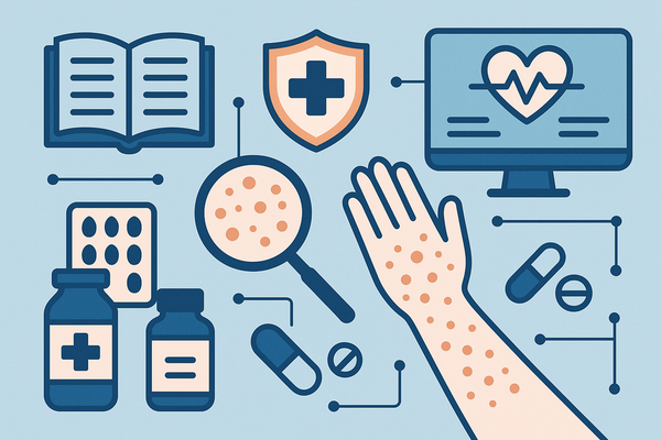
Section 1: Overview of AI in Rash Detection
Keywords: AI rash detection, machine learning, computer vision, deep learning
AI rash detection combines several core technologies:
- Machine learning:
- Models trained on labeled skin images detect visual patterns.
- Performance improves as more data and annotations accumulate.
- Deep learning & Convolutional Neural Networks (CNNs):
- Multi-layer architectures automatically extract hierarchical features (edges, textures, color).
- CNNs excel at segmentation (isolating rash areas) and classification (categorizing rash types).
- Computer vision:
- Algorithms for feature detection identify lesion borders, color distribution, and texture.
- Enables automated image-based assessment and computer-aided diagnosis.
Current trends and advancements include:
- Expanding beyond pigmented lesion classification to cover infectious and inflammatory rashes.
- Multimodal models that fuse clinical metadata (age, symptoms) with images for richer context.
- Open-source datasets and federated learning frameworks that preserve privacy by training across decentralized data sources.
Key benefits of AI rash detection:
- Rapid throughput: processes hundreds of images in seconds vs. minutes for manual review.
- Objectivity and reproducibility: standardized assessments reduce inter-observer variability.
- Improved access: remote clinics and underserved regions gain diagnostic support, cutting time-to-diagnosis.
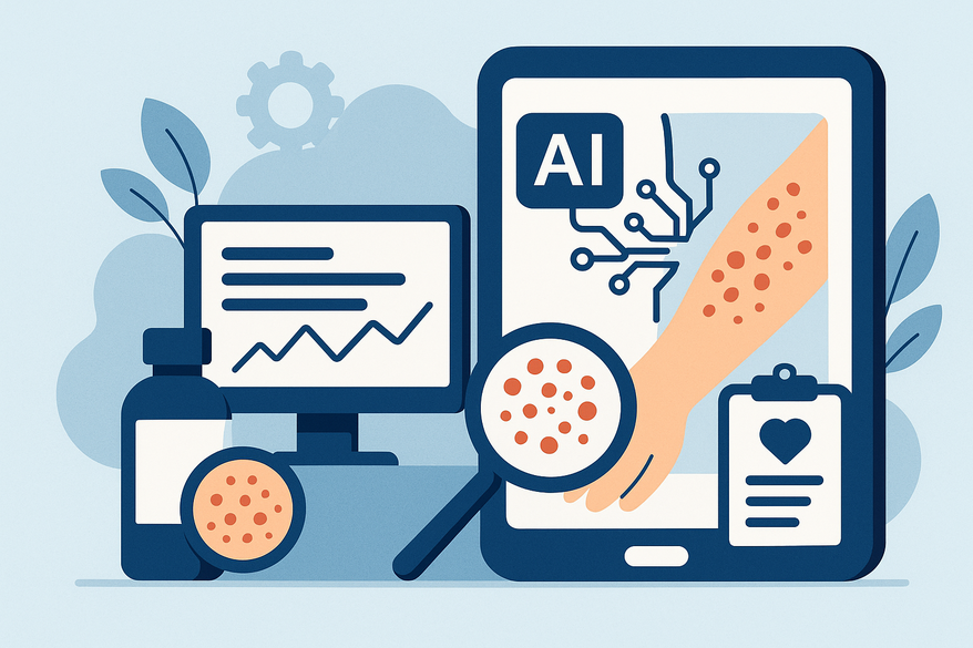
Clinicians and patients can also leverage Rash Detector, an AI skin analysis app that generates instant sample reports for images of rashes and skin issues.
Section 2: Investigating Real World Effectiveness
Keywords: real world effectiveness of AI rash detection, practical effectiveness, clinical implementation
Case Study A: Pigmented Lesion Classification
• Finding: Machine-learning classifiers achieved 91.8% accuracy vs. 89.8% for dermatologists (2.01% absolute improvement).
• Metrics: sensitivity (malignant detection rate), specificity (benign exclusion), overall accuracy.
• Implication: AI can match or exceed expert performance on high-quality images.
Case Study B: GP-Powered Image Analysis Model
• Prospective validation with general practitioners analyzing patient rashes.
• Top-1 accuracy: 31.7%; Top-5 accuracy: 68.1%.
• Implication: Top-N predictions support triage—narrowing differential diagnoses—while definitive decisions remain clinician-led.
Case Study C: YOLO v4 & Multimodal Neural Networks
• Integration of clinical metadata boosted sensitivity from 82% to 89%, specificity from 78% to 85% for infectious rashes.
• Deployed in outpatient dermatology clinics for real-time decision support.
• Impact: Faster lesion screening and more consistent triage recommendations.
Synthesis of Findings:
- Accuracy ranges: 70–92% depending on condition and dataset.
- Common errors: false positives on benign variations; false negatives on early-stage or atypical presentations.
- Throughput: up to 500 images/hour with cloud-based inference pipelines.
- Variability: performance shifts with dataset diversity, image quality, and workflow integration.
For more on reliability and performance metrics, see how accurate rash detection AI
Section 3: Challenges and Limitations
Keywords: limitations, challenges, data quality
Variation in Skin Tones
• Underrepresentation of darker Fitzpatrick skin types leads to lower sensitivity on pigmented lesions and inflammatory rashes.
• Disparities: sensitivity drops by up to 15% on skin types V–VI vs. I–III in some models.
Atypical Rash Presentations
• Non-standard morphology—mixed lesions, overlapping conditions—confuse pattern recognition algorithms.
• Examples: morbilliform drug eruptions vs. varicella-like vesicular rashes can share color and texture features, triggering misclassification.
Data Quality Issues
• Factors: image resolution, inconsistent lighting, varied anatomical sites.
• Impact: reduced model confidence and reproducibility when images deviate from training distribution.
Error Types
• False positives: benign eczema flagged as psoriasis or infection, generating unnecessary follow-ups.
• False negatives: missing early malignant changes or rare conditions, delaying treatment.
• Comparison: dermatologist error rates vary 5–10% based on experience level, highlighting that AI is a complement, not a replacement.
Ethical, Privacy, and Regulatory Considerations
• Data protection: HIPAA (US) and GDPR (EU) require secure handling of patient images and metadata.
• Regulatory clearance: FDA (USA) and CE marking (EU) demand clinical validation trials and transparency of algorithm performance.
• Informed consent: patients must understand AI’s role and limitations when used in diagnosis.
Further discussion of these limitations is available at limitations of AI rash detection in dermatology
Section 4: Comparison with Traditional Methods
Keywords: comparison, traditional dermatology, real world effectiveness of AI rash detection
- Accuracy: AI rash detection: comparable or higher accuracy for specific lesions (e.g., 92% vs. 90% dermatologist accuracy on pigmented nevi). Traditional dermatology: accuracy depends heavily on clinician expertise—residents vs. board-certified specialists.
- Efficiency: AI: analyzes an image in 1–2 seconds, enabling batch processing and real-time triage. Traditional: in-person exam requires 10–15 minutes per patient, including history and physical exam.
- Cost: AI: significant upfront R&D and deployment costs, followed by low marginal cost per case. Traditional: ongoing personnel costs, clinic overhead, and patient travel expenses.
- Scalability & Access: AI: supports teledermatology platforms, 24/7 triage, and remote screening programs in rural areas. Traditional: limited by clinic hours, geographic specialist shortages, and patient wait times.
Defining Real World Effectiveness Metrics:
Sensitive metrics: true positive and true negative rates in routine practice, positive and negative predictive values reflecting prevalence, time-to-diagnosis, and cost-per-case as operational metrics for health systems.
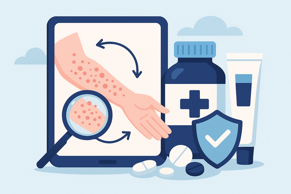
Section 5: Future of AI Rash Detection, Innovations, Teledermatology
Keywords: future of AI rash detection, innovations, teledermatology
- Enhancing Model Generalizability: Diverse datasets including Fitzpatrick types I–VI to reduce bias; federated learning for privacy-preserving collaboration.
- Teledermatology Integration: Mobile apps and remote clinics with embedded AI triage modules; augmented intelligence assists clinicians while retaining final decision authority.
- Personalized Medicine & Monitoring: AI-driven treatment recommendations leveraging patient history, genomics, and comorbidities; automated tracking of lesion progression via smartphone images.
- Regulatory Frameworks & Expert Consensus: Upcoming guidelines on validation protocols, performance thresholds, and post-market surveillance to integrate AI tools as standard adjuncts in dermatology.
Conclusion
The real world effectiveness of AI rash detection is now supported by multiple clinical studies and pilot programs. AI matches or exceeds traditional accuracy for specific lesion types, processes images much faster, and expands access through teledermatology. However, challenges—skin tone biases, data quality variability, ethical and regulatory hurdles—must be addressed through diverse data collection, federated learning, transparent validation, and robust governance.
AI holds immense promise for accelerating diagnosis, bridging specialist shortages, and improving patient outcomes. Clinicians and administrators should:
- Stay informed about emerging evidence and regulatory updates
- Pilot AI tools in controlled practice settings
- Contribute to data diversity efforts (e.g., annotating underrepresented skin types)
By collaborating across disciplines—dermatology, data science, regulatory bodies—we can realize the full potential of AI rash detection to improve care equitably and sustainably.
FAQ
- How accurate is AI rash detection in clinical practice?
AI models demonstrate 70–92% accuracy depending on the condition and dataset; with high-quality images and diverse training data, accuracy can match or exceed expert dermatologists. - Can AI tools identify rashes on all skin tones?
Models currently underrepresent darker Fitzpatrick skin types, leading to sensitivity gaps. Diverse datasets and federated learning aim to improve performance across all skin tones. - Is patient data protected when using AI apps?
Reputable platforms comply with HIPAA and GDPR, use encryption, and often employ federated learning to process data locally, minimizing privacy risks. - Will AI replace dermatologists?
AI serves as an adjunct, providing rapid, standardized assessments. Final diagnoses and treatment decisions remain with clinicians to ensure holistic patient care.